38 cell diagram and labels
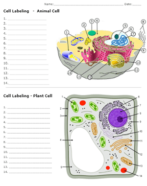
animal and plant cell diagram to label - TeachersPayTeachers Animal & Plant Cell: Label the Diagram and Differences table:This is a great supplement for students to review/assess and strengthen their knowledge the unit of ANIMAL AND PLANT CELL UNIT. Answer key included.It includes total TWO worksheets. This worksheets can demonstrates relationships between facts and concepts.It can be used as a small ... Crypto Goes to Washington | Time Oct 03, 2022 · The inter-agency pissing match is the subject of endless speculation and argument among crypto people, but it’s important less in its particulars than what it signifies: would-be crypto ...
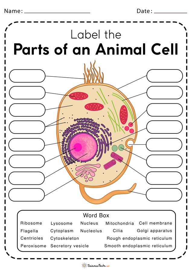
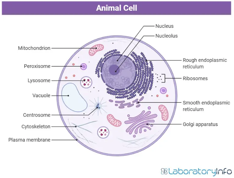
Animal Cell- Definition, Structure, Parts, Functions, Labeled Diagram An animal cell is a eukaryotic cell that lacks a cell wall, and it is enclosed by the plasma membrane. The cell organelles are enclosed by the plasma membrane including the cell nucleus. Unlike the animal cell lacking the cell wall, plant cells have a cell wall. Animals are a large group of diverse living organisms that make up three-quarters ...

Cell diagram and labels
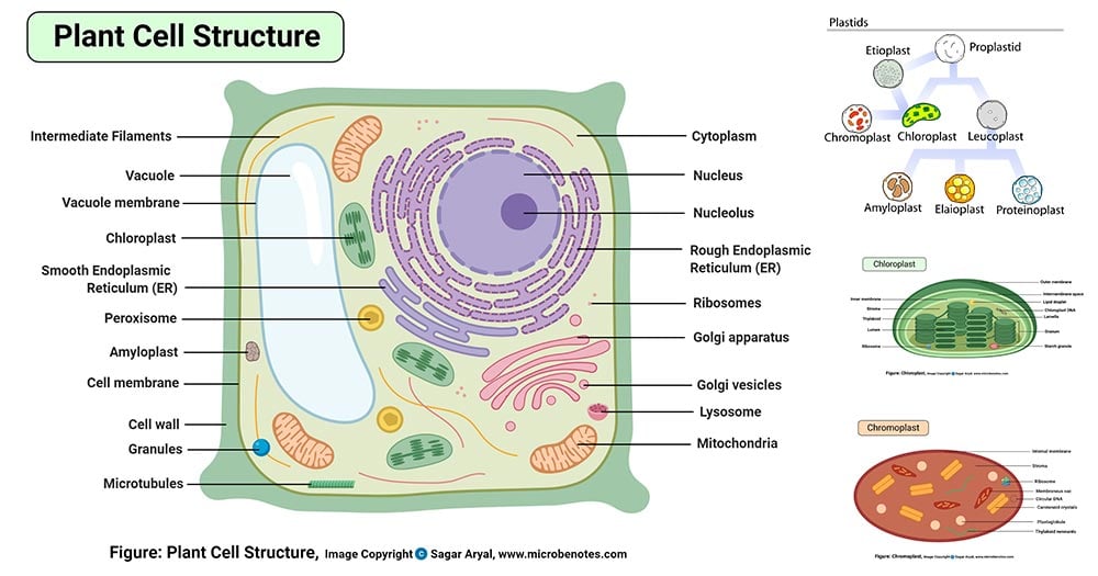
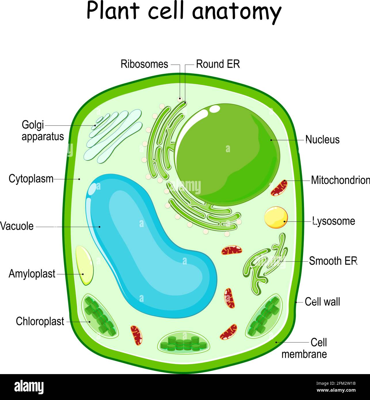
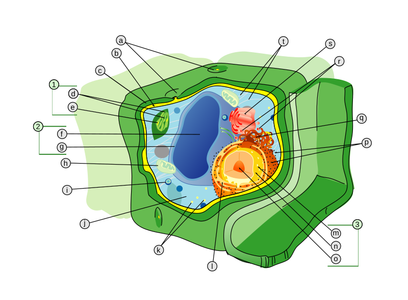
Plant Cell: Diagram, Types and Functions - Embibe Exams These differences can be clearly understood when the cells are examined under an electron microscope. Observe the labelled diagram of plant cell structure as given below: Are Plant Cells Prokaryotic or Eukaryotic? The cell is the basic structural and functional unit of life in all living organisms. Structure of Cell: Definition, Cell Theory, Plant and Animal Cells - Embibe Various kinds of cells show special differences, yet they all have some basic structural plan consisting of three essential parts: (i) cell membrane ( plasma membrane ), (ii) cytoplasm and (iii) nucleus. Apart from these three components, cells have some living parts that are called cell organelles. Animal and Plant Cell Worksheets - Super Teacher Worksheets This is a basic illustration of a plant cell with major parts labeled. Labels include nucleus, chloroplast, cytoplasm, membrane, cell wall, and vacuole, and mitochondrion. Use it as a poster in your classroom or have students glue it into their science notebooks.
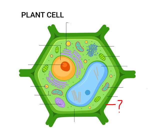
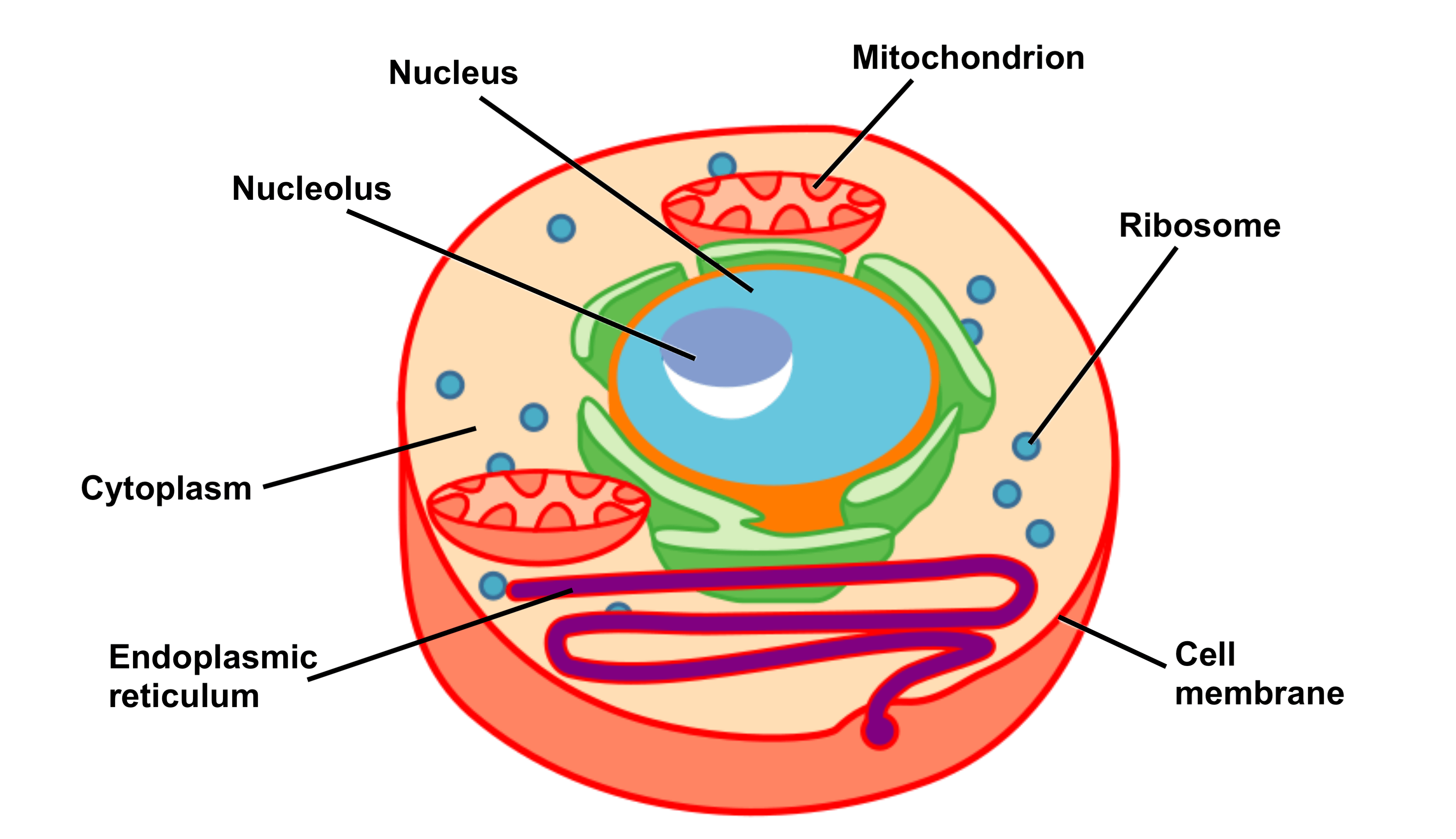
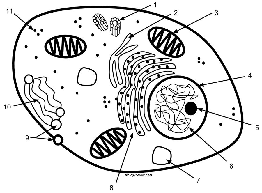
Cell diagram and labels. Plant Cell Diagram | Science Trends A plant cell diagram, like the one above, shows each part of the plant cell including the chloroplast, cell wall, plasma membrane, nucleus, mitochondria, ribosomes, etc. A plant cell diagram is a great way to learn the different components of the cell for your upcoming exam. Plants are able to do something animals can't: photosynthesize. Animal Cells: Labelled Diagram, Definitions, and Structure - Research Tweet The endoplasmic reticulum (s) are organelles that create a network of membranes that transport substances around the cell. They have phospholipid bilayers. There are two types of ER: the rough ER, and the smooth ER. The rough endoplasmic reticulum is rough because it has ribosomes (which is explained below) attached to it. Web Content Accessibility Guidelines (WCAG) 2.0 - W3 Dec 11, 2008 · Abstract. Web Content Accessibility Guidelines (WCAG) 2.0 covers a wide range of recommendations for making Web content more accessible. Following these guidelines will make content accessible to a wider range of people with disabilities, including blindness and low vision, deafness and hearing loss, learning disabilities, cognitive limitations, limited movement, speech disabilities ... A Labelled Diagram Of Neuron with Detailed Explanations - BYJUS A Labelled Diagram Of Neuron with Detailed Explanations Biology Biology Article Diagram Of Neuron Diagram Of Neuron A neuron is a specialized cell, primarily involved in transmitting information through electrical and chemical signals. They are found in the brain, spinal cord and the peripheral nerves. A neuron is also known as the nerve cell.
03 Label the Cell Diagram | Quizlet Start studying 03 Label the Cell. Learn vocabulary, terms, and more with flashcards, games, and other study tools. ... cell diagram. 18 terms. lugo_janet. Sets found in the same folder. 03 Organelle Functions. 14 terms. muskopf1. 07 Cell Labeling. 11 terms. muskopf1. 03 Cell Transport. 12 terms. A Labeled Diagram of the Animal Cell and its Organelles A Labeled Diagram of the Animal Cell and its Organelles There are two types of cells - Prokaryotic and Eucaryotic. Eukaryotic cells are larger, more complex, and have evolved more recently than prokaryotes. Where, prokaryotes are just bacteria and archaea, eukaryotes are literally everything else. A Well-labelled Diagram Of Animal Cell With Explanation - BYJUS The animal cell diagram is widely asked in Class 10 and 12 examinations and is beneficial to understand the structure and functions of an animal. A brief explanation of the different parts of an animal cell along with a well-labelled diagram is mentioned below for reference. Also Read Different between Plant Cell and Animal Cell Learn the parts of a cell with diagrams and cell quizzes Cell diagram unlabeled It's time to label the cell yourself! As you fill in the cell structure worksheet, remember the functions of each part of the cell that you learned in the video. Doing this will help you to remember where each part is located. Click the links below to download the labeled and unlabeled eukaryotic cell diagrams.
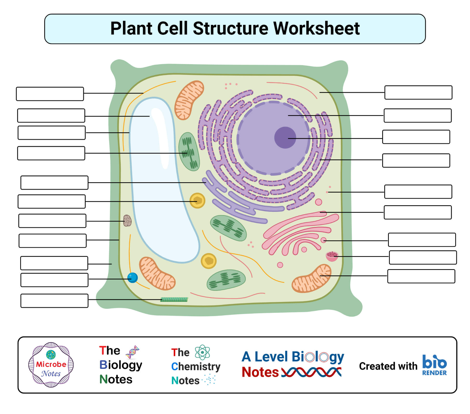
Converting Diagrams - The Biology Corner Open Google Draw and import the diagram. Then use "insert" to create text boxes where students can fill in the labels. Don't forget when assigning this to students on Google classroom to make a copy for each student. You can leave documents in an uneditable form and students can use an addon like "Kami" to annotate the document. A Labeled Diagram of the Plant Cell and Functions of its Organelles ... The cell membrane is a thin layer made up of proteins, lipids, and fats. It forms a protective wall around the organelles contained within the cell. It is selectively permeable and thus, regulates the transportation of materials needed for the survival of the organelles of the cell. Function: Protects the cell from its surroundings. Labeling a Cell Diagram | Quizlet Cell Wall This gives shape and support to the plant cell. It surrounds the cell and protects the other parts of the cell. Chloroplasts This is where the plant cell's chlorophyll is stored. This is what the plant uses to make its own food (photosynthesis). This is also what makes plant cells have a green-like color. Plant cells Are circular in shape Could Call of Duty doom the Activision Blizzard deal? - Protocol Oct 14, 2022 · A MESSAGE FROM QUALCOMM Every great tech product that you rely on each day, from the smartphone in your pocket to your music streaming service and navigational system in the car, shares one important thing: part of its innovative design is protected by intellectual property (IP) laws.
Animal Cell Diagram & Anatomy - Enchanted Learning
Cell Organelles- Definition, Structure, Functions, Diagram Cilia and Flagella are tiny hair-like projections from the cell made of microtubules and covered by the plasma membrane. Structure of Cilia and Flagella Cilia are hair-like projections that have a 9+2 arrangement of microtubules with a radial pattern of 9 outer microtubule doublet that surrounds two singlet microtubules.
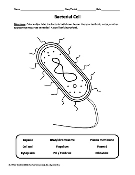
Bacterial Cell Labeling Diagram
Interactive Cell Model - CELLS alive Cell Wall. Chloroplast. Smooth Endoplasmic Reticulum. Rough Endoplasmic Reticulum. Ribosomes. Cytoskeleton. RETURN to CELL DIAGRAM ...
Plant Cell- Definition, Structure, Parts, Functions, Labeled ...
Label the cell diagram - Teaching resources - Wordwall Label the cell diagram Examples from our community 10000+ results for 'label the cell diagram' label Cell Cycle diagram Labelled diagram by Alecbaus El desayuno (label the diagram) Labelled diagram by Betsyjansey G9 Spanish El desayuno Label the Cell Membrane Labelled diagram by Renatathomas Label the Plant Cell Labelled diagram by Armstrem

GCSE Biology Cell Structure Labelling Diagrams | Teaching ...
Cell Anatomy - Labeling Animal and Plant Cells - YouTube Learn the cell structures and how to identify and label them.Like. Share. Subscribe.
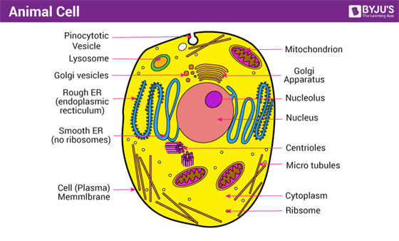
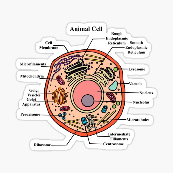
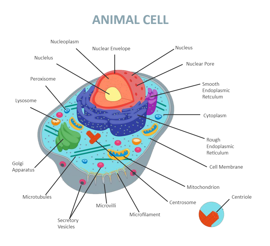
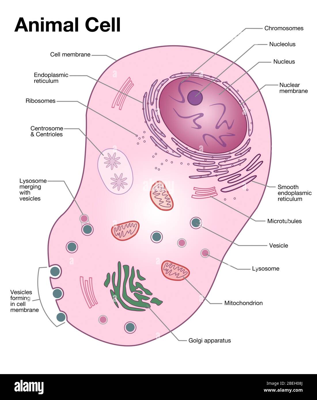
A Well-labelled Diagram Of Animal Cell With Explanation
141,670 Labelled Cell Images, Stock Photos & Vectors | Shutterstock Find Labelled cell stock images in HD and millions of other royalty-free stock photos, illustrations and vectors in the Shutterstock collection. Thousands of new, high-quality pictures added every day.
Most People Can't Identify 10/12 Of The Parts Of A Cell — Can ...
Picture of the Ear: Ear Conditions and Treatments - WebMD WebMD's Ear Anatomy Page provides a detailed image and definition of the ear as well as an overview of ear-related health problems. Learn about the ear's function in the body and test and ...
Animal Cell Worksheets - Free Printable
Structure and mechanism of human cystine exporter cystinosin Sep 15, 2022 · Removing the NTD reduced cystine transport activity by ∼70% in a cell-based uptake assay (Figure 2C) and severely impaired the expression and stability of cystinosin (Figures S4H and S4J), suggesting an important structural role. This is consistent with early discoveries that an internal deletion in the NTD is linked to cystinosis and causes ...

Animal Cell - The Definitive Guide | Biology Dictionary
Cell Labeling Quiz - PurposeGames.com This is an online quiz called Cell Labeling. There is a printable worksheet available for download here so you can take the quiz with pen and paper.
Labeled Animal Cell Diagram Sticker by BundaBear
Flow cytometry - Wikipedia Flow cytometry (FC) is a technique used to detect and measure physical and chemical characteristics of a population of cells or particles.. In this process, a sample containing cells or particles is suspended in a fluid and injected into the flow cytometer instrument. The sample is focused to ideally flow one cell at a time through a laser beam, where the light scattered is …

THE HUMAN CELL. | Cell, Human, Map
Mathematical formulation of the Standard Model - Wikipedia The diagram shows the elementary particles of the Standard Model (the Higgs boson, the three generations of quarks and leptons, and the gauge bosons), including their names, masses, spins, charges, chiralities, and interactions with the strong, weak and electromagnetic forces.
Cell Structure and Function Part 1 – The Organelles - Medical ...
Cell - Label | Cell Structure Quiz - Quizizz Play this game to review Cell Structure. Label #3

Interactive cell diagram by Diann Caviness
Cells Diagram | Science Illustration Solutions - Edrawsoft Cells Diagram Symbols Edraw software offers you lots of symbols used in cells diagram like cell structure, paramecium, squamous cell, cell division, bacteria, cell membrane, eggs, sperm, zygote, an animal cell, SARS, tobacco mosaic, adenovirus, coliphage, herpesvirus, AIDS, pollen, plant cell model, onion tissue, etc. Cells Diagram Examples
Label Cell Parts | Plant & Animal Cell Activity | StoryboardThat
PDF Human Cell Diagram, Parts, Pictures, Structure and Functions Structure and Functions The cell is the basic functional in a human meaning that it is a self-contained and fully operational living entity. Humans are multicellular organisms with various different types of cells that work together to sustain life. Other non-cellular components in the body include water, macronutrients

Cell Structure
Free Cell Diagram Software with Free Templates - EdrawMax - Edrawsoft An animal cell diagram describes a cell structure enclosed by a plasma member, and it has a nucleus with a membrane and organelles. Neuron Diagram A neuron diagram describes the three parts of a Neuron: dendrites, an axon, a cell body, or soma. Cell Membrane Diagram

Label the Cell Diagram | Quizlet
Label the cell - Teaching resources - Wordwall Label Plant and Animal Cell Labelled diagram by Eawilson 5.6 Label the sentence Labelled diagram by Christianjolene Label the Electromagnetic Spectrum Labelled diagram by Elizabetheck G6 G7 G8 Science 5.7 Label the sentence Labelled diagram by Christianjolene the cell Match up by Elenagp9149 8.1 Label the sentence Labelled diagram
Plant cell structure. vector diagram. anatomy of a biological ...
label a animal cell Cell Structure Diagrams. 16 Images about Cell Structure Diagrams : Label Animal Cell - Improve your science knowledge with free questions in animal cell diagrams, cell label - Dr. Hunter Biology and also Animal Cell Structure Without Labels - Structure Of Animal And Plant Cell Download Scientific. Cell Structure Diagrams mandevillehigh.stpsb.org

Label a cell, Labeling parts of a cell, Cells Structures and ...
Cell: Structure and Functions (With Diagram) - Biology Discussion Eukaryotic Cells: 1. Eukaryotes are sophisticated cells with a well defined nucleus and cell organelles. 2. The cells are comparatively larger in size (10-100 μm). 3. Unicellular to multicellular in nature and evolved ~1 billion years ago. 4. The cell membrane is semipermeable and flexible. 5. These cells reproduce both asexually and sexually.

The Animal and Plant Cells Colour and Label Diagram ...
Labeled Plant Cell With Diagrams | Science Trends The parts of a plant cell include the cell wall, the cell membrane, the cytoskeleton or cytoplasm, the nucleus, the Golgi body, the mitochondria, the peroxisome's, the vacuoles, ribosomes, and the endoplasmic reticulum. Parts Of A Plant Cell The Cell Wall Let's start from the outside and work our way inwards.

Cell Structure Label Organ Cell Stock Vector (Royalty Free ...
Human Cell Diagram, Parts, Pictures, Structure and Functions Diagram of the human cell illustrating the different parts of the cell. Cell Membrane. The cell membrane is the outer coating of the cell and contains the cytoplasm, substances within it and the organelle. It is a double-layered membrane composed of proteins and lipids. The lipid molecules on the outer and inner part (lipid bilayer) allow it to ...
Label the Parts of the Plant and Animal Cell
Animal and Plant Cell Worksheets - Super Teacher Worksheets This is a basic illustration of a plant cell with major parts labeled. Labels include nucleus, chloroplast, cytoplasm, membrane, cell wall, and vacuole, and mitochondrion. Use it as a poster in your classroom or have students glue it into their science notebooks.

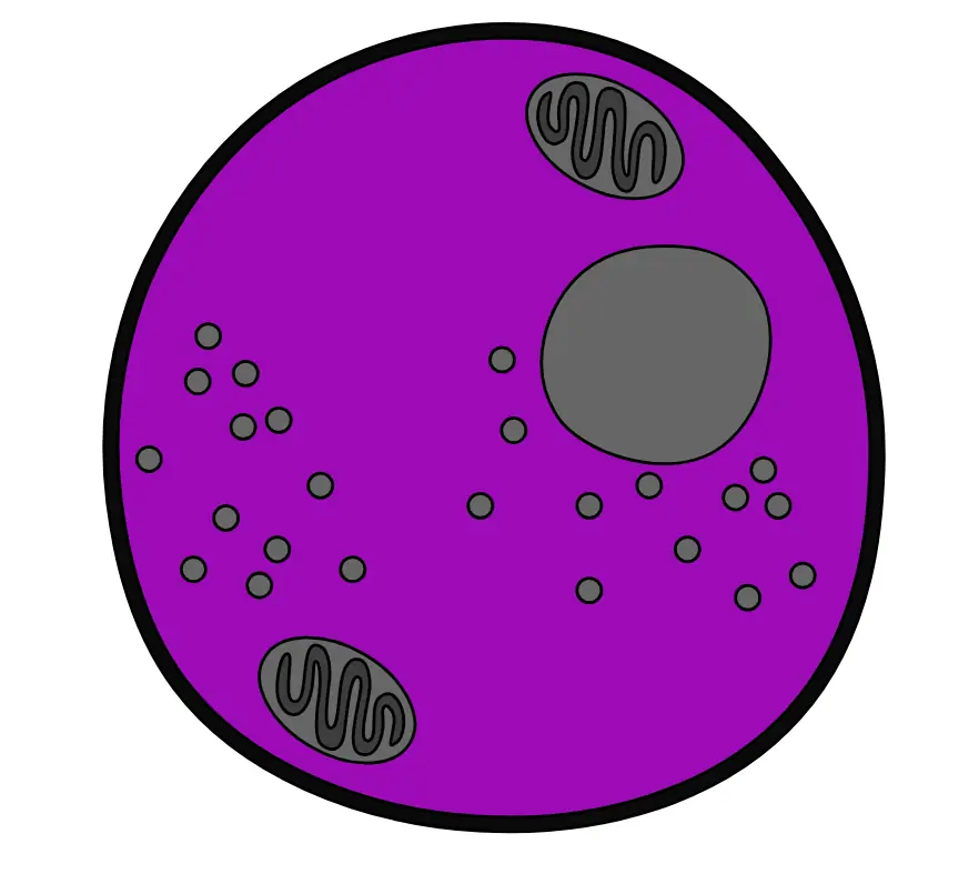
Printable Animal Cell Diagram – Labeled, Unlabeled, and Blank ...
Structure of Cell: Definition, Cell Theory, Plant and Animal Cells - Embibe Various kinds of cells show special differences, yet they all have some basic structural plan consisting of three essential parts: (i) cell membrane ( plasma membrane ), (ii) cytoplasm and (iii) nucleus. Apart from these three components, cells have some living parts that are called cell organelles.

Animal Cell - Free printable to label + Color | Célula animal ...
Plant Cell: Diagram, Types and Functions - Embibe Exams These differences can be clearly understood when the cells are examined under an electron microscope. Observe the labelled diagram of plant cell structure as given below: Are Plant Cells Prokaryotic or Eukaryotic? The cell is the basic structural and functional unit of life in all living organisms.
Plant Cell- Definition, Structure, Parts, Functions, Labeled ...

cell structure | Cell diagram, Human cell diagram, Animal ...
Cell wall - Wikipedia
(434).jpg)
Animal Cell Labeling Quiz Questions And Answers - ProProfs Quiz
Cell Label - Simple vs. Complex

A Labeled Diagram of the Animal Cell and its Organelles ...
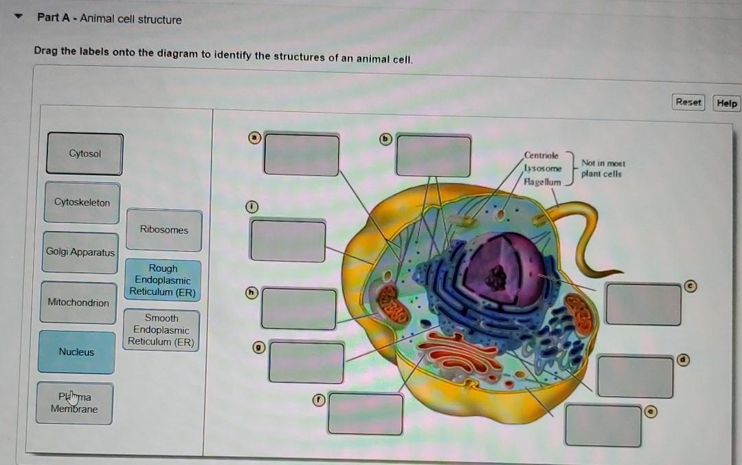
Solved Part A - Animal cell structure Drag the labels onto ...

Animal Cell- Definition, Structure, Parts, Functions, Labeled ...
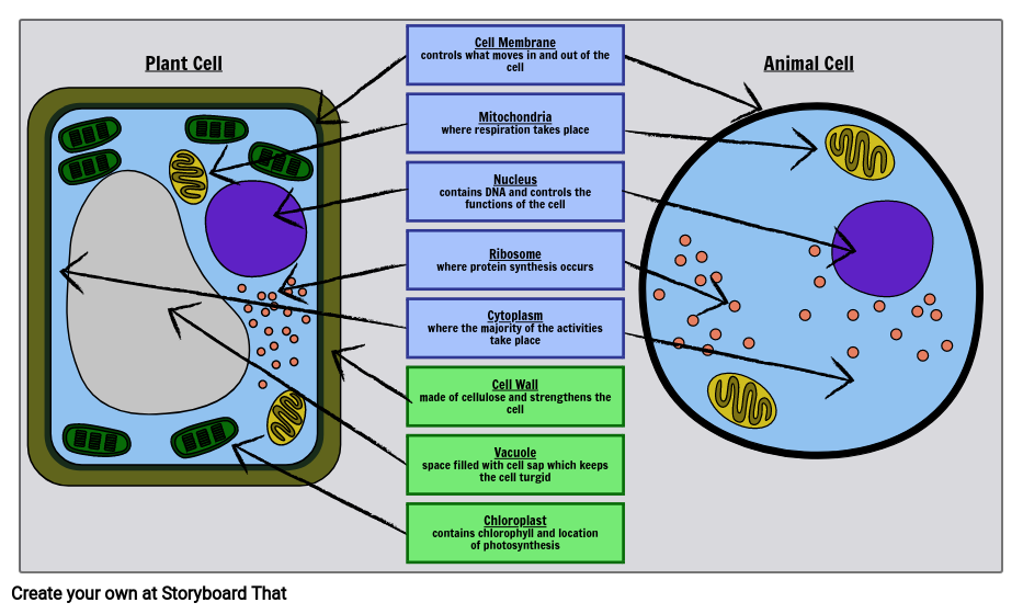
Label Cell Parts | Plant & Animal Cell Activity | StoryboardThat
Animal Cell Diagram Labeled free image download
Animal Cell Diagram Labeled | EdrawMax Template
Detailed guide on Animal Cell and its parts (with labelled ...

4,503 Human Cell Diagram Stock Photos, Pictures & Royalty ...
Animal cell diagram hi-res stock photography and images - Alamy
Animal cell - Teaching resources

Eukaryotic Cells | BioNinja
Post a Comment for "38 cell diagram and labels"