44 cell structure with labels
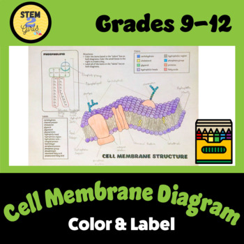
› entEntertainment & Arts - Los Angeles Times L.A. Times entertainment news from Hollywood including event coverage, celebrity gossip and deals. med.libretexts.org › Bookshelves › Anatomy_and4.1: Cell Structure and Function - Medicine LibreTexts Aug 13, 2020 · Glycoproteins and glycolipids in the membrane act as identification markers or labels on the extracellular surface of the membrane. Thus, the plasma membrane has many functions and works as both a gateway and a selective barrier. Figure \(\PageIndex{2}\) Phospholipids form the basic structure of a cell membrane. Hydrophobic tails of ...
Label a cell, Labeling parts of a cell, Cells Structures and Functions ... cell membrane. Regulates what enters or leaves the cell. cell wall. A rigid outer layer that surrounds plant cells, supports and protects the cell. large central vacuoles. the source of water for the cell. Chloroplast. Where photosynthesis occurs to make oxygen and glucose, only found in plant cells. Ribosome.
Cell structure with labels
corner.bigblueinteractive.com › indexThe Corner Forum - New York Giants Fans Discussion Board ... Big Blue Interactive's Corner Forum is one of the premiere New York Giants fan-run message boards. Join the discussion about your favorite team! Bacteria in Microbiology - shapes, structure and diagram - Jotscroll Bacteria cells are the smallest living cells that are known; even though viruses are smaller than bacteria, viruses are not living cells. In microbiology there are different types of bacteria with various sizes, shapes, and structures. The bacteria shapes, structure, and labeled diagrams are discussed below. Label the cell structure. | Homework.Study.com Eukaryotic cells, including human cells, contain a nucleus (with the exception of red blood cells) and organelles. Organelles are membrane-enclosed structures ...
Cell structure with labels. Label Cell Parts | Plant & Animal Cell Activity | StoryboardThat Create a cell diagram with each part of plant and animal cells labeled. Include descriptions of what each organelle does. Click "Start Assignment". Find diagrams of a plant and an animal cell in the Science tab. Using arrows and Textables, label each part of the cell and describe its function. Animal Cell Structure Labeling Game - PurposeGames.com This is an online quiz called Animal Cell Structure Labeling Game There is a printable worksheet available for download here so you can take the quiz with pen and paper. From the quiz author Match the name of each membrane bound organelle to the correct structure in the cell. Your Skills & Rank Total Points 0 Get started! Today's Rank -- 0 Cell Organelles - Types, Structure and their Functions - BYJUS Ribosomes are found in the form of tiny particles in a large number of cells and are mainly composed of 2/3rd of RNA and 1/3rd of protein. They are named as the 70s (found in prokaryotes) or 80s (found in eukaryotes) The letter S refers to the density and the size, known as Svedberg's Unit. Both 70S and 80S ribosomes are composed of two subunits. Draw a diagram of a plant cell and label at least eight class 11 ... Hint: Plant cell has cell wall whereas cell wall is absent in an animal cell. The chloroplast is present only in plant cells. Plant cells contain large ...
Cell Diagrams with Labelling Activity - Learnful The cell structure illustrations for these diagrams were generated in BioRender. Both diagrams feature a drag-and-drop labelling activity created with H5P ... Label animal cell - Teaching resources - Wordwall 10000+ results for 'label animal cell'. Label Animal Cell Organelles Labelled diagram. by Britter. Label Animal Cell Organelles Labelled diagram. by Mbauer. Label Plant and Animal Cell Labelled diagram. by Catherine34. Animal Cell Label Labelled diagram. by Taraabbott. Cell Organelles- Definition, Structure, Functions, Diagram - Microbe Notes In a plant cell, the cell wall is made up of cellulose, hemicellulose, and proteins while in a fungal cell, it is composed of chitin. A cell wall is multilayered with a middle lamina, a primary cell wall, and a secondary cell wall. The middle lamina contains polysaccharides that provide adhesion and allow binding of the cells to one another. Cell - Label | Cell Structure Quiz - Quizizz 16 Questions Show answers Question 1 30 seconds Q. Label #3 answer choices Nucleus Mitochondria Vacuole Golgi Body Question 2 30 seconds Q. Label #4 answer choices Cell wall Cell membrane Nuclear membrane Gatekeeper Question 3 30 seconds Q. Label #5 answer choices Nucleus Nucleolus Endoplasmic Reticulum Mitochondria Question 4 30 seconds
In cell a what is the structure labeled x? Explained by FAQ Blog On average, the cells in your body are replaced every 7 to 10 years. But those numbers hide a huge variability in lifespan across the different organs of the body. Neutrophil cells (a type of white blood cell) might only last two days, while the cells in the middle of your eye lenses will last your entire life. Labeled Plant Cell With Diagrams | Science Trends The parts of a plant cell include the cell wall, the cell membrane, the cytoskeleton or cytoplasm, the nucleus, the Golgi body, the mitochondria, the peroxisome's, the vacuoles, ribosomes, and the endoplasmic reticulum. Parts Of A Plant Cell The Cell Wall Let's start from the outside and work our way inwards. Structure of Cell: Definition, Types, Diagram, Functions - Embibe Cells are the fundamental structural and functional unit of all living beings including plants, animals and microorganisms. All living organisms in this universe are made up of cells. We cannot see cells with naked eyes as they are only \ (10\) microns in size whereas human eyes cannot see objects less than \ (100\) microns. Plant Cell- Definition, Structure, Parts, Functions, Labeled Diagram Plant cells are eukaryotic cells, that are found in green plants, photosynthetic eukaryotes of the kingdom Plantae which means they have a membrane-bound nucleus. They have a variety of membrane-bound cell organelles that perform various specific functions to maintain the normal functioning of the plant cell. Structure of Plant cell
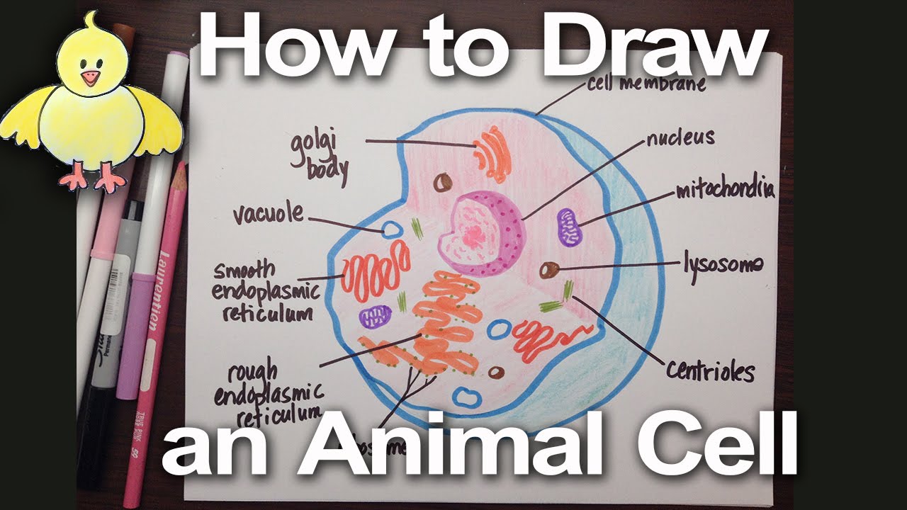
How to Draw an Animal Cell Diagram -Homework Help | DoodleDrawArt
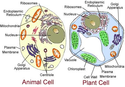
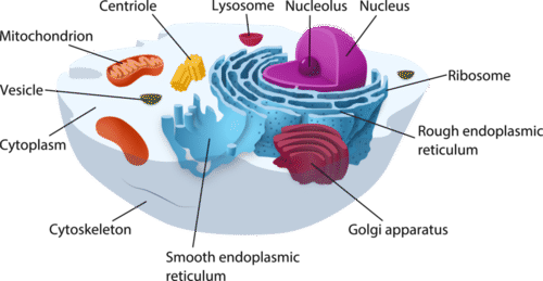
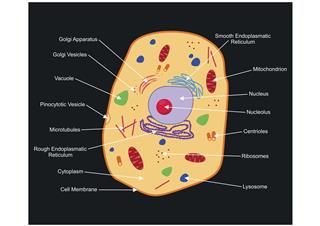
A Labeled Diagram of the Animal Cell and its Organelles A Labeled Diagram of the Animal Cell and its Organelles There are two types of cells - Prokaryotic and Eucaryotic. Eukaryotic cells are larger, more complex, and have evolved more recently than prokaryotes. Where, prokaryotes are just bacteria and archaea, eukaryotes are literally everything else.
Label animal cell - Teaching resources
Draw a diagram of a typical cell and label the following class 11 ... Hint: Cell is the basic structural and functional unit of all organisms. It is the smallest unit of life. They are generally known as the building blocks of ...

07 Cell Labeling Flashcards | Quizlet
Cell Structure | Thermo Fisher Scientific - US Cell structure labels can be a counterstain method to identify the location of specific proteins and targets of interest within the cell. Our cell labels include cell structure specific dyes or antibodies for live-cell or fixed-cell imaging, as well as for flow cytometry. Cell structure protocols Cytoskeleton Structure
Cell Structure
Eukaryotic Cell: Definition, Structure & Function (with Analogy ... Eukaryotic cells include animal cells - including human cells - plant cells, fungal cells and algae. Eukaryotic cells are characterized by a membrane-bound nucleus. That's distinct from prokaryotic cells, which have a nucleoid - a region that's dense with cellular DNA - but don't actually have a separate membrane-bound compartment like the nucleus.

CELLS Blank Plant & Animal Cell Diagrams: Note Taking ...
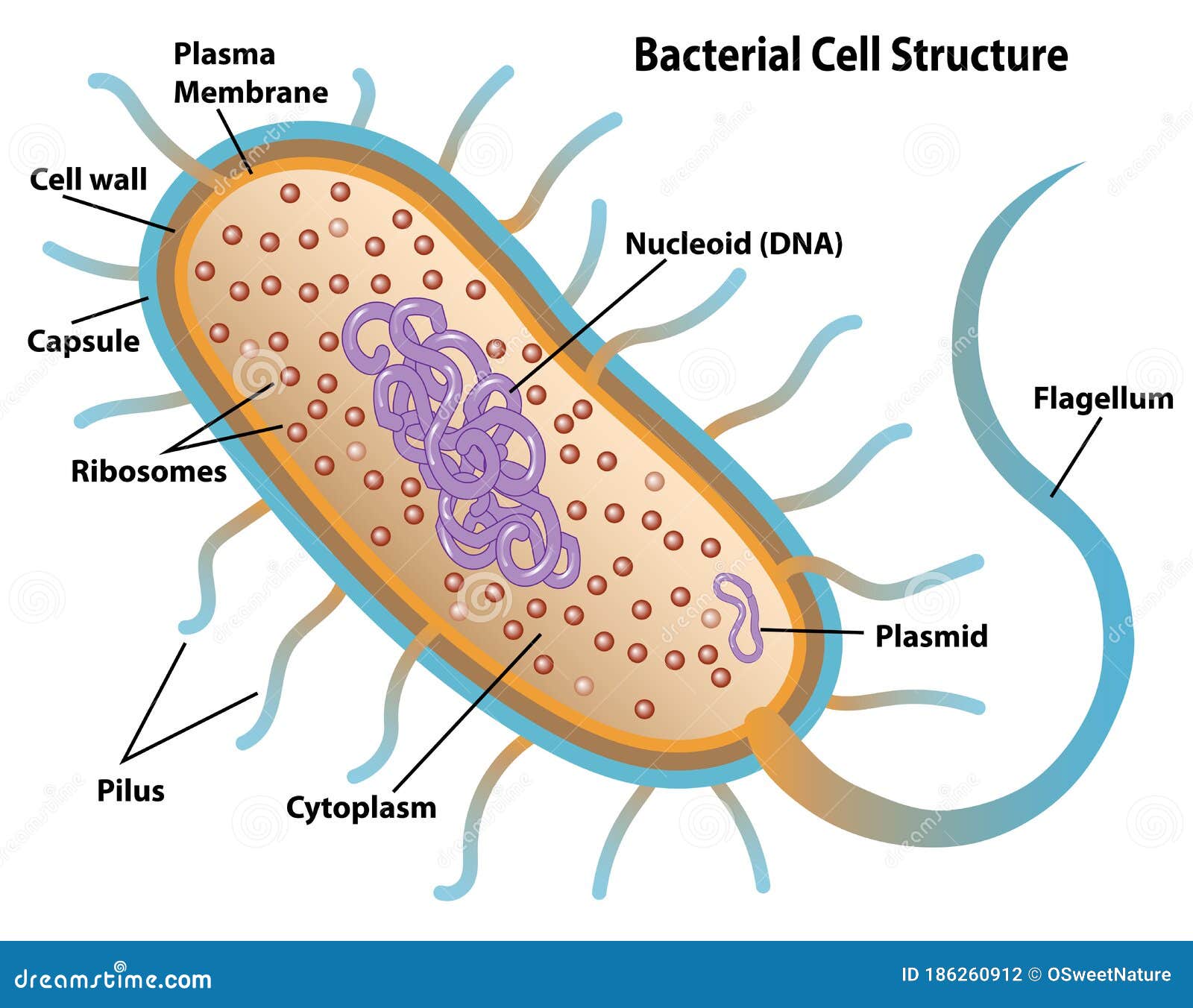
Bacteria Cell Structures with labels - Dreamstime Bacteria Cell Structures with labels Royalty-Free Vector Bacterial cell structures labeled on a bacillus cell with nucleoid DNA and ribosomes. External structures include the capsule, pili, and flagellum. Morphology of internal structures of bacteria. cell anatomy bacteria, prokaryotic cell, cell, internal structures, prokaryotic, dna, bacteria,
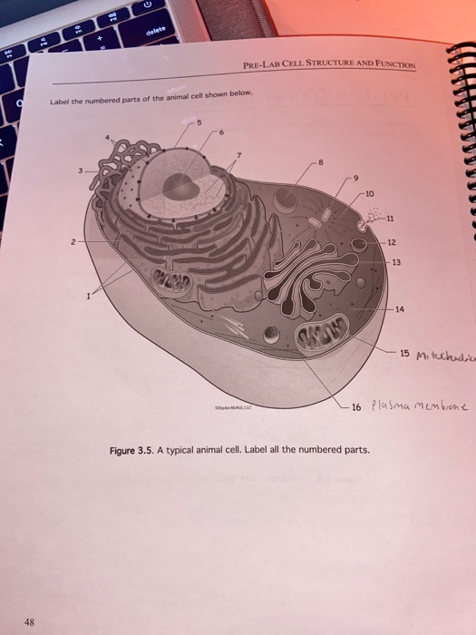
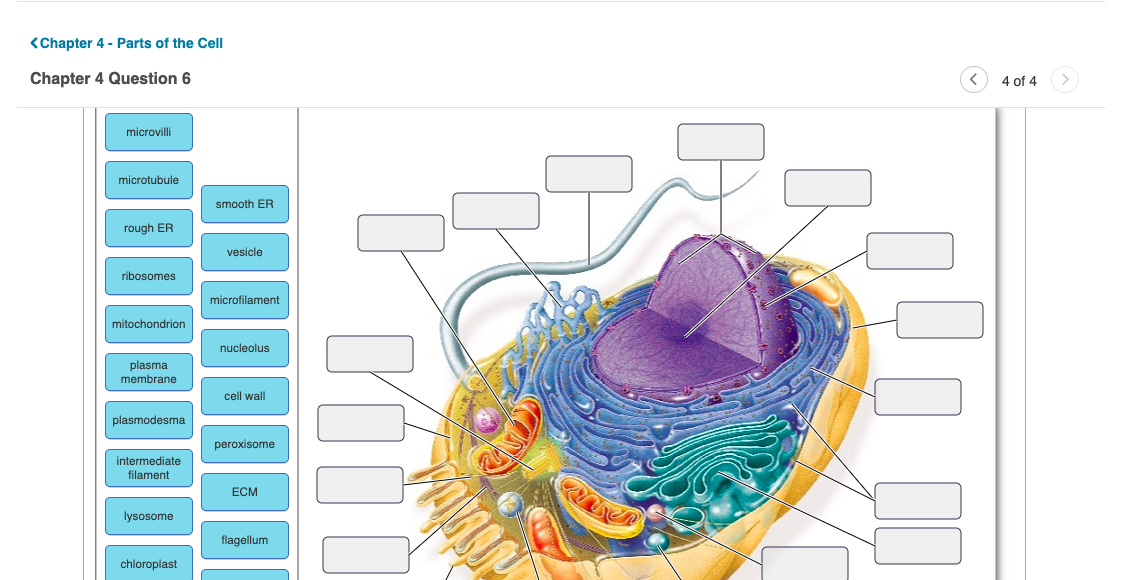
delete P LAR CELL STRUCTURE AND FUNCTION Label the | Chegg.com
Get Label That Diagram - Cells from the Microsoft Store This app provides the user the opportunity to study cell structure as well as test what they know. There are 5 cells presented: Animal Cell, Plant Cell, Amoeba, Paramecium and a Euglena. The player can study the labeled diagram or play the game of labeling the diagram. When the game is played, the labels appear in a random order one at a time and the player must tap on the correct dot on the ...

Eukaryotic Cell Structure Labeling: Animal Diagram | Quizlet
Human Cell Diagram, Parts, Pictures, Structure and Functions The cell is the basic functional in a human meaning that it is a self-contained and fully operational living entity. Humans are multicellular organisms with various different types of cells that work together to sustain life. ... The endoplasmic reticulum (ER) is a membranous structure that contains a network of tubules and vesicles. Its ...

15 Cell - Structure ideas | cell structure, cell, animal cell
Cell: Structure and Functions (With Diagram) - Biology Discussion Eukaryotic Cells: 1. Eukaryotes are sophisticated cells with a well defined nucleus and cell organelles. 2. The cells are comparatively larger in size (10-100 μm). 3. Unicellular to multicellular in nature and evolved ~1 billion years ago. 4. The cell membrane is semipermeable and flexible. 5. These cells reproduce both asexually and sexually.
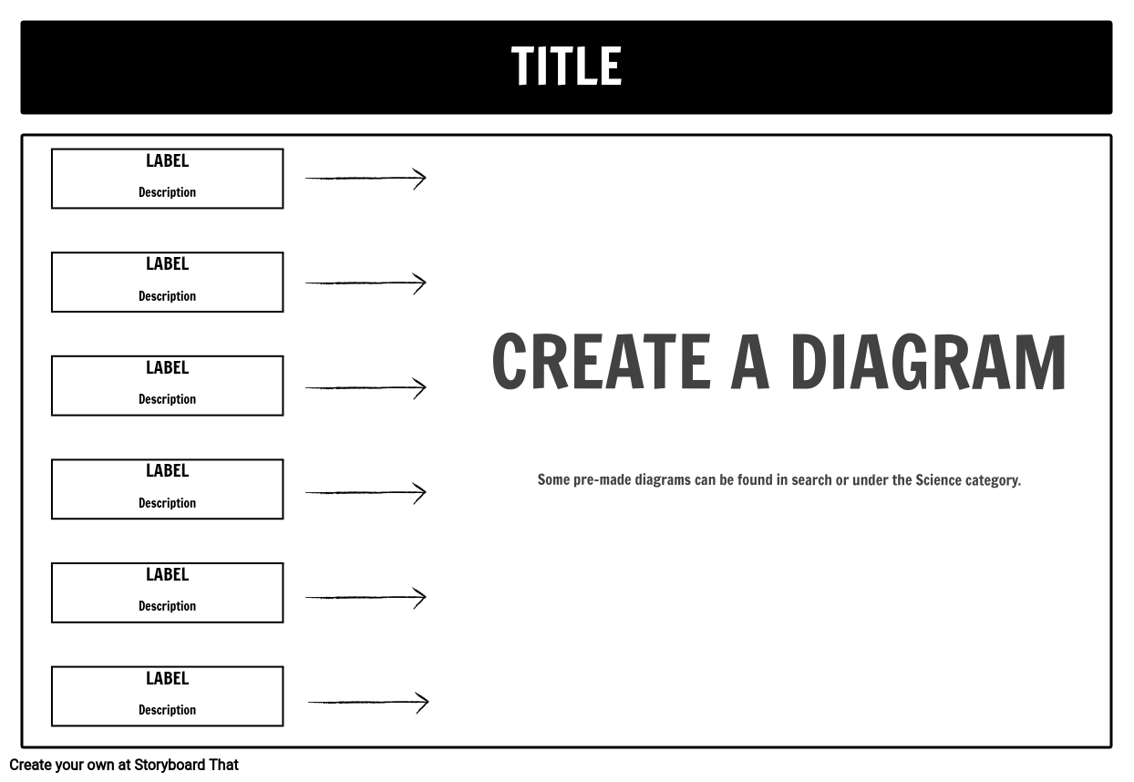
Label Cell Parts | Plant & Animal Cell Activity | StoryboardThat
cell structure | Cell diagram, Human cell diagram, Animal cell drawing structures shown here will seldom all be found in a single animal cell ... Free printable cell worksheets for coloring pages, label the cell, notebooking, ...
Cell Structure Label Teaching Resources | Teachers Pay Teachers
Label the cell structure. | Homework.Study.com Label the cell structure. Cells: All living cells contain an intracellular space called the cytoplasm. The cytoplasm is filled with a jelly-like fluid where many of the cells enzymatic reactions...

Cell Structure
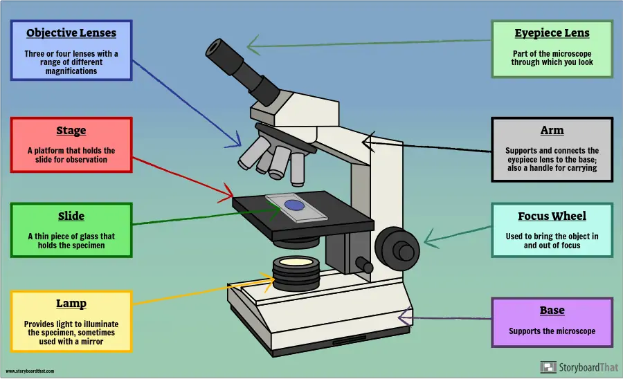
› bitesize › guidesAnimal cells - Cell structure - Edexcel - GCSE Combined ... A generalised animal cell and its components. Mitochondria (singular: mitochondrion) are visible with a light microscope but can't be seen in detail. Ribosomes are only visible with an electron ...
Label Cell Parts | Plant & Animal Cell Activity | StoryboardThat
THE HUMAN CELL. | Cell, Human, Biology - Pinterest Feb 15, 2021 - THE QUESTION TO THIS IS: Correctly label the following structures in a typical human cell.
Cell Structures | CK-12 Foundation
Cell Structure of Bacteria (With Diagram) - Biology Discussion In this article we will discuss about the cell structure of bacteria with the help of diagrams. A bacterial cell (Fig. 2.5) shows a typical prokaryotic structure. The cytoplasm is enclosed by three layers, the outermost slime or capsule, the middle cell wall and inner cell membrane. The major cytoplasmic contents are nucleoid, plasmid, ribosome ...
Solved Label the structures of the animal cell below. Drag ...
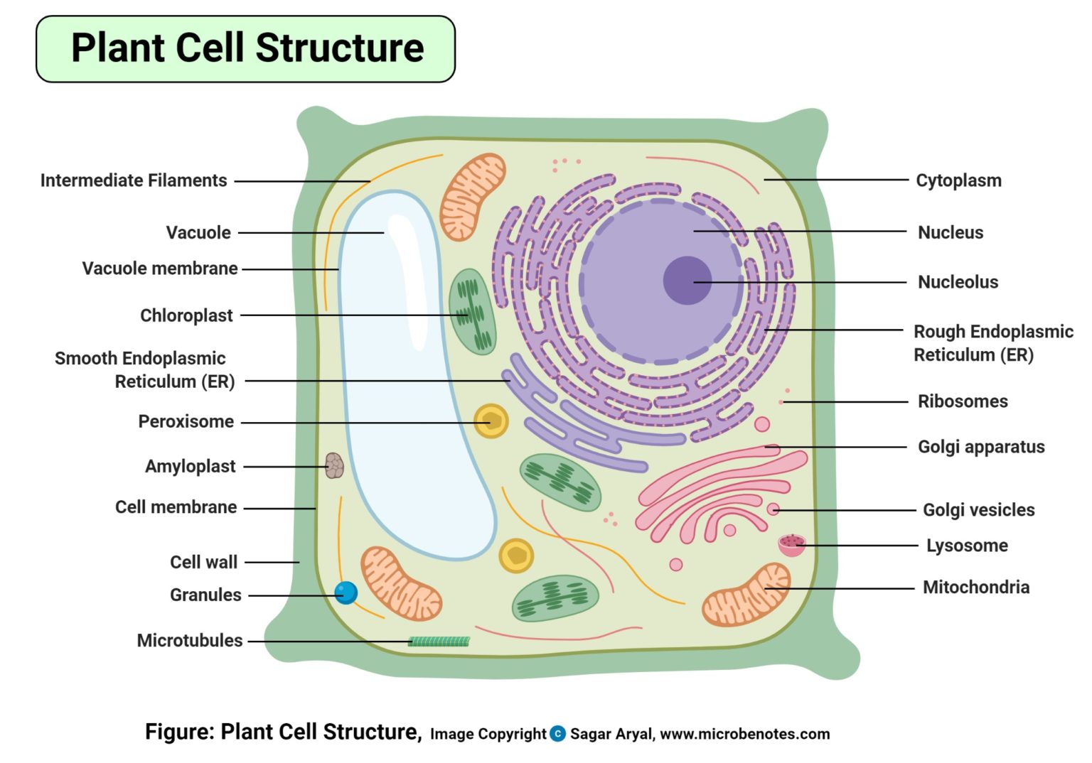
Plant Cell - Definition, Structure, Function, Diagram & Types - BYJUS It is a rigid layer which is composed of polysaccharides cellulose, pectin and hemicellulose. It is located outside the cell membrane. It also comprises glycoproteins and polymers such as lignin, cutin, or suberin. The primary function of the cell wall is to protect and provide structural support to the cell.

File:Plant cell structure svg labels.svg - Wikimedia Commons
Cell Structure | Thermo Fisher Scientific - FR Cell structure labels can be a counterstain method to identify the location of specific proteins and targets of interest within the cell. Our cell labels ...

Animal Cell - Free printable to label + Color | Célula animal ...
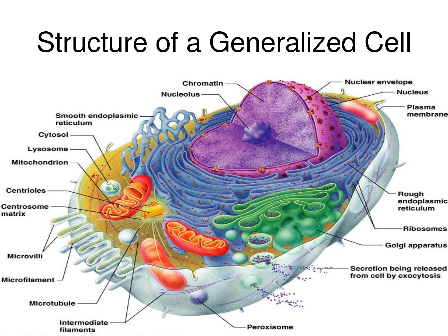
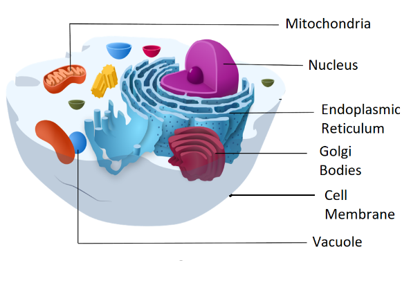
Cell Structure - SEER Training It includes features from all cell types. A cell consists of three parts: the cell membrane, the nucleus, and, between the two, the cytoplasm. Within the ...

Label the Animal Cell Worksheets (SB11866) | Animal cells ...
Cell Structure and Function Quiz - mrskerrett.com Cell Structure and Function Quiz ... Label 2 is . Label 3 is . Label 4 is . Label 5 is Label 6 is . Label 7 is . Label 8 is . Label 9 is . label 10 is 11. This structure is made of DNA . 12. Produces ATP . 13. Creates turgor pressure . 14. New proteins are ...
Cell Diagram With Structure and Function | A Definitive Guide
141374 Labelled Cell Images, Stock Photos & Vectors - Shutterstock Cell organelles biological vector illustration diagram. ... Bacterial cell anatomy labeling structures on a bacillus cell with nucleoid DNA and ribosomes.

Cell Organelles and Function with Labels Flashcards | Quizlet
› TR › html4Forms in HTML documents - W3 17.9 Labels. Some form controls automatically have labels associated with them (press buttons) while most do not (text fields, checkboxes and radio buttons, and menus). For those controls that have implicit labels, user agents should use the value of the value attribute as the label string.

Cell Structure Label Teaching Resources | Teachers Pay Teachers
› cell › fulltextMapping information-rich genotype-phenotype landscapes ... - Cell Jun 09, 2022 · Mapping the relationship between genetic changes and their phenotypic consequence is critical to understanding gene and cellular function. This mapping is traditionally carried out in one of two ways: a phenotype-centric, “forward genetic” approach that reveals the genetic changes that drive a phenotype of interest or a gene-centric, “reverse genetic” approach that catalogs the diverse ...

Printable Animal Cell Diagram – Labeled, Unlabeled, and Blank
Plant Cells: Labelled Diagram, Definitions, and Structure - Research Tweet Plastids and Chloroplasts. Plants make their own food through photosynthesis. Plant cells have plastids, which animal cells don't. Plastids are organelles used to make and store needed compounds. Chloroplasts are the most important of plastids. They convert light energy from the sun into sugar and oxygen. The most exposed parts of the plants ...

Eukaryotic Cells | BioNinja
en.wikipedia.org › wiki › Building_insulation_materialBuilding insulation material - Wikipedia Building insulation materials are the building materials which form the thermal envelope of a building or otherwise reduce heat transfer.. Insulation may be categorized by its composition (natural or synthetic materials), form (batts, blankets, loose-fill, spray foam, and panels), structural contribution (insulating concrete forms, structured panels, and straw bales), functional mode ...

Eukaryote Structure | BioNinja
Animal Cells: Labelled Diagram, Definitions, and Structure - Research Tweet The endoplasmic reticulum (s) are organelles that create a network of membranes that transport substances around the cell. They have phospholipid bilayers. There are two types of ER: the rough ER, and the smooth ER. The rough endoplasmic reticulum is rough because it has ribosomes (which is explained below) attached to it.

Labelled Diagram Of A Human Cell Bone Cell Labeled Diagram ...
Plant Cell-Definition, Structure, Parts, Functions, Labeled Diagram Structure of Plant cell. Typically, plant cells are bigger than animal cells of equivalent size and structure. Typically, they have a cubic or rectangular shape. ... Figure: Labeled diagram of a plant cell, created with biorender.com. The plant cell is comprised of cellulose, hemicellulose, and pectin, as well as plastids, which are essential ...
Learn the parts of a cell with diagrams and cell quizzes | Kenhub
03 Label the Cell Diagram | Quizlet Start studying 03 Label the Cell. Learn vocabulary, terms, and more with flashcards, games, and other study tools.

Animal Cells | Animal cell, Cell model, Cell biology
Plant Cell: Meaning, Components, Structure, Functions & Parts - Embibe The structures t are as follows: Plant Cell Wall It is a rigid layer that is composed of cellulose, glycoproteins, lignin, pectin and hemicellulose. It is located outside the cell membrane and is completely permeable.
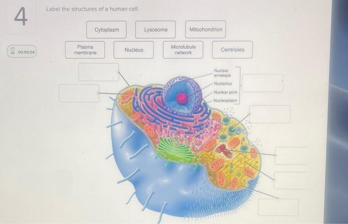
Solved Label the structures of a human cell. 4 Cytoplasm ...
Plant Cell Structure and Parts Explained With a Labeled Diagram Different Parts of a Plant Cell. Plant cells are classified into three types, based on the structure and function, viz. parenchyma, collenchyma and sclerenchyma. The parenchyma cells are living, thin-walled and undergo repeated cell division for growth of the plant. They are mostly present in the leaf epidermis, stem pith, root and fruit pulp.

BIO101 – Cell Structure | A Blog Around The Clock
GCSE Biology Cell Structure Labelling Diagrams | Teaching ... 17 Oct 2021 ...

How can I label the diagram and know if it is a plant or ...
Label the cell structure. | Homework.Study.com Eukaryotic cells, including human cells, contain a nucleus (with the exception of red blood cells) and organelles. Organelles are membrane-enclosed structures ...
Solved
Bacteria in Microbiology - shapes, structure and diagram - Jotscroll Bacteria cells are the smallest living cells that are known; even though viruses are smaller than bacteria, viruses are not living cells. In microbiology there are different types of bacteria with various sizes, shapes, and structures. The bacteria shapes, structure, and labeled diagrams are discussed below.

Cell Structure Label Organ Cell Stock Vector (Royalty Free ...
corner.bigblueinteractive.com › indexThe Corner Forum - New York Giants Fans Discussion Board ... Big Blue Interactive's Corner Forum is one of the premiere New York Giants fan-run message boards. Join the discussion about your favorite team!

Animal Cell Diagram To Label - Biology Forums Gallery
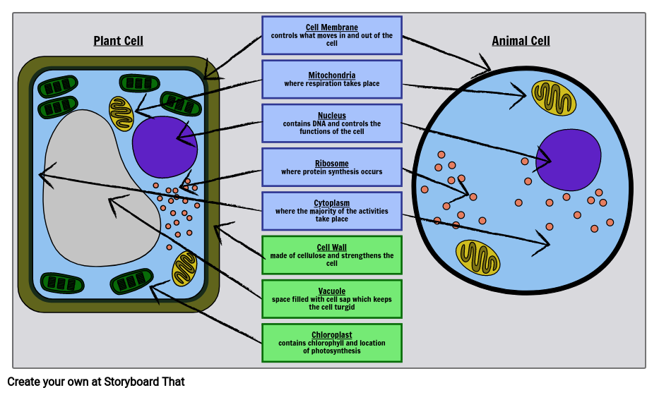
Label Cell Parts | Plant & Animal Cell Activity | StoryboardThat

Lab Manual Exercise # 1a

Cell Structure and Function Quiz
Bacteria Cell Structures with Labels Stock Vector ...
A Labeled Diagram of the Animal Cell and its Organelles ...
Draw a diagram of a typical cell and label the following ...
Plant Cell- Definition, Structure, Parts, Functions, Labeled ...
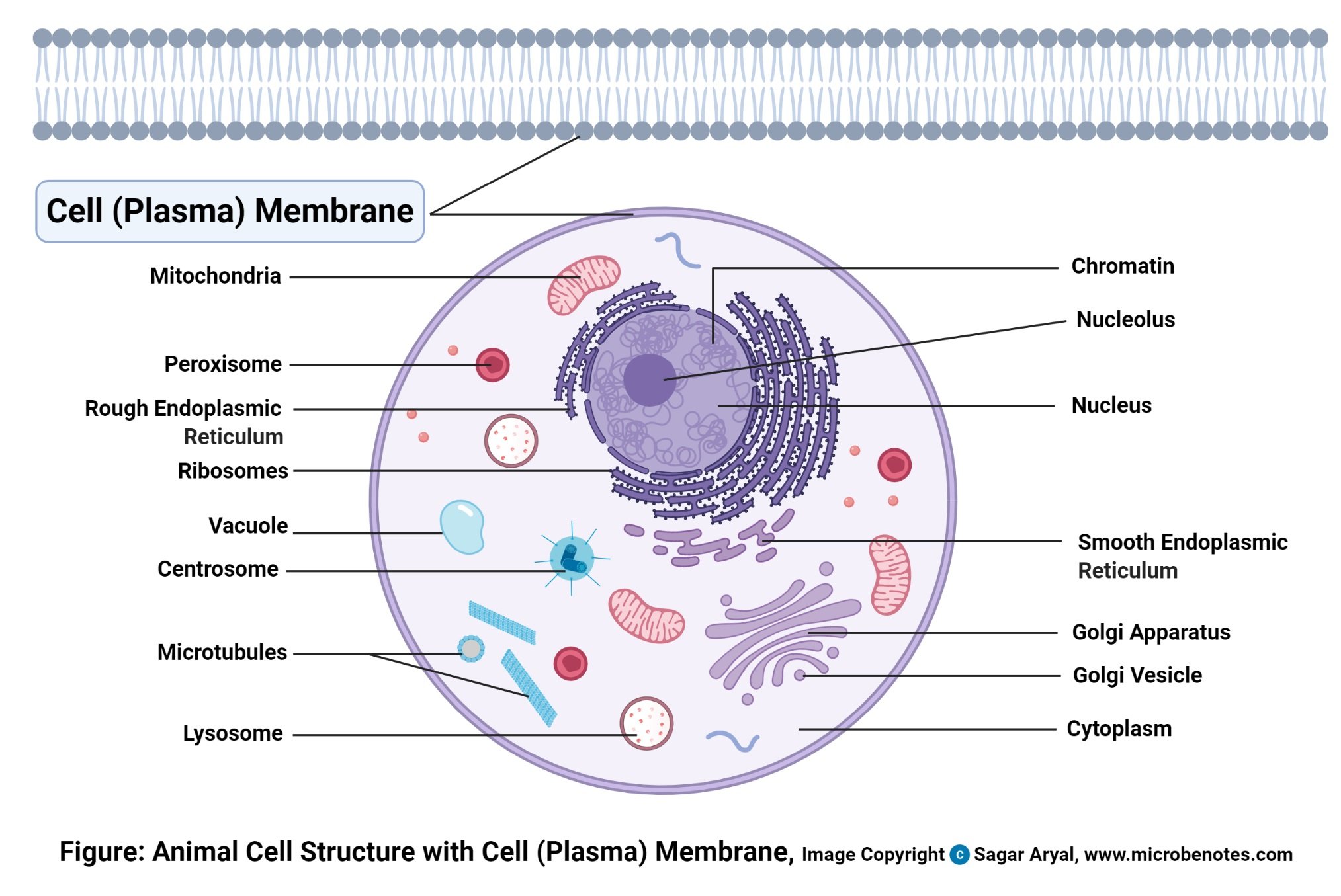
Animal Cell- Definition, Structure, Parts, Functions, Labeled ...
cell | Definition, Types, Functions, Diagram, Division ...
SEER Training: Cell Structure

Cell Structure Label Organ Cell Stock Vector (Royalty Free ...

Printable Animal Cell Diagram – Labeled, Unlabeled, and Blank
Post a Comment for "44 cell structure with labels"