43 diagram of a cell with labels
How to draw an animal cell - labeled science diagram - YouTube Download a free printable outline of this video and draw along with us: you for watching. Please ... Cell Diagrams - The Biology Corner Open Google Draw and import the diagram. Then use "insert" to create text boxes where students can fill in the labels. Don't forget when assigning this to students on Google classroom to make a copy for each student. You can leave documents in an uneditable form and students can use an addon like "Kami" to annotate the document.
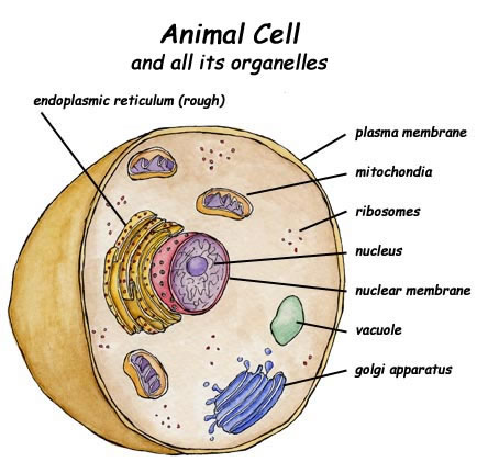
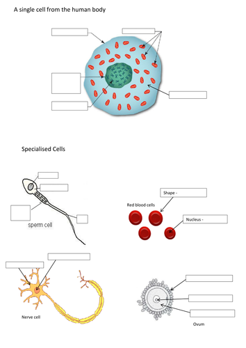
Animal Cell Diagram | Science Trends The diagram, like the one above, will include labels of the major parts of an animal cell including the cell membrane, nucleus, ribosomes, mitochondria, vesicles, and cytosol. The cells of animals are the basic structural units for the wide variety of life we see in the animal kingdom. Animal cells are eukaryotic in nature, possessing a nucleus ...

Diagram of a cell with labels
Label the cell - Teaching resources - Wordwall G5 Science. Label the Plant Cell Labelled diagram. by Koneal2. G7 Biology. Label Animal Cell Organelles Labelled diagram. by Britter. Correctly Label the Bacteria (Prokaryotic) Cell Labelled diagram. by Bronwyn12. Label Plant and Animal Cell Labelled diagram. Diagram of a cell membrane with labels - NIST Essential Biological FunctionsImmune response, Cell metabolism, Neurotransmission, Photosynthesis, Cell adherence, Cell growth and differentiationPotential Commercial ApplicationsDrug response monitoring, Chemical manufacturing, Biosensing, Energy conversion, Tissue engineering ... Diagram of a cell membrane with labels. Appears In. Biology in ... Cell Organelles- Definition, Structure, Functions, Diagram Cell organelles are specialized entities present inside a particular type of cell that performs a specific function. There are various cell organelles, out of which, some are common in most types of cells like cell membranes, nucleus, and cytoplasm. However, some organelles are specific to one particular type of cell-like plastids and cell ...
Diagram of a cell with labels. Animal Cells: Labelled Diagram, Definitions, and Structure Animal Cells Organelles and Functions. A double layer that supports and protects the cell. Allows materials in and out. The control center of the cell. Nucleus contains majority of cell's the DNA. Popularly known as the "Powerhouse". Breaks down food to produce energy in the form of ATP. 03 Label the Cell Diagram | Quizlet Start studying 03 Label the Cell. Learn vocabulary, terms, and more with flashcards, games, and other study tools. PDF Human Cell Diagram, Parts, Pictures, Structure and Functions Diagram of the human cell illustrating the different parts of the cell. Cell Membrane The cell membraneis the outer coating of the cell and contains the cytoplasm, substances within it and the organelle. It is a double-layered membrane composed of proteins and lipids. A Labeled Diagram of the Plant Cell and Functions of its Organelles A Labeled Diagram of the Plant Cell and Functions of its Organelles We are aware that all life stems from a single cell, and that the cell is the most basic unit of all living organisms. The cell being the smallest unit of life, is akin to a tiny room which houses several organs. Here, let's study the plant cell in detail...
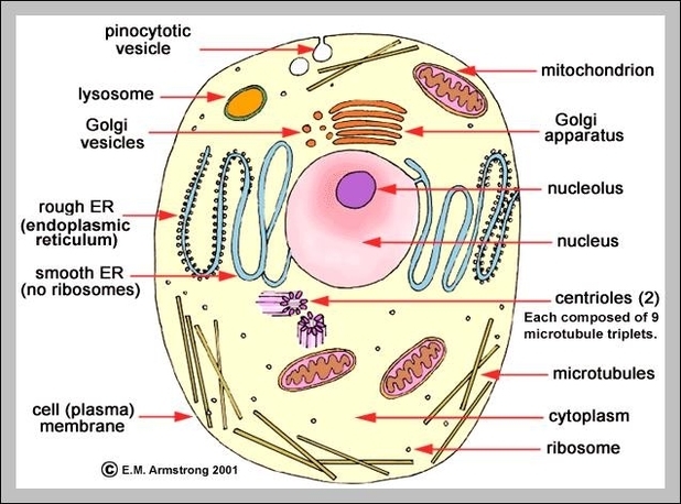
Drawing & Labeling a Diagram of a Electrochemical Cell - Study.com Lesson Summary. An electrochemical cell consists of two half-cells connected by a salt bridge.Each half-cell consist of a metal strip in a solution of that metal. Oxidation occurs in one half-cell ... diagram of a cell labeled diagram of a cell labeled Paramecium under 400x magnification. Explain the nucleus of a cell with a neat labeled diagram. Volvox cells protist colony flagella practical lab daughter quia parent amoeba feldman 1b bio vocabulary diagram of a cell labeled A Well-labelled Diagram Of Animal Cell With Explanation - Byju's Well-Labelled Diagram of Animal Cell The Cell Organelles are membrane-bound, present within the cells. There are various organelles present within the cell and are classified into three categories based on the presence or absence of membrane. Listed below are the Cell Organelles of an animal cell along with their functions. Examine the diagram of a cell. Which accurately labels the lysosome? W ... Explanation: Lysosomes are heterogeneous structures present in animal cells which are bound by single membranes. They are of varying shape and size and contains hydrolytic enzymes inside it. Lysosomal membrane has H⁺ ATPase which pumps H⁺ into the membrane through ATP hydrolysis. This pumping of H⁺ makes the internal pH of lysosome acidic.
Label Cell Parts | Plant & Animal Cell Activity | StoryboardThat Create a cell diagram with each part of plant and animal cells labeled. Include descriptions of what each organelle does. Click "Start Assignment". Find diagrams of a plant and an animal cell in the Science tab. Using arrows and Textables, label each part of the cell and describe its function. A Labeled Diagram of the Animal Cell and its Organelles One can observe the golgi apparatus in the labeled animal cell parts diagram. The golgi apparatus is situated near the cell nucleus and besides the stacked sacs, it also contains large number of vesicles. The main function of this golgi complex is to receive the proteins synthesized in the ER and transform it into more complex proteins. Labeled Plant Cell With Diagrams | Science Trends The parts of a plant cell include the cell wall, the cell membrane, the cytoskeleton or cytoplasm, the nucleus, the Golgi body, the mitochondria, the peroxisome's, the vacuoles, ribosomes, and the endoplasmic reticulum. Parts Of A Plant Cell The Cell Wall Let's start from the outside and work our way inwards. Animal Cell Diagram with Label and Explanation: Cell Structure, Functions Diagram of Animal Cell Below is the diagram of the animal cell which shows the organelles present in it. The cell is covered with cytoplasm which consists of cell organelles in it. The nucleus is covered with a rough Endoplasmic Reticulum and other organelles each designed for a specific purpose.
cell diagrams to label | animal cell (diagram & label)(7-2) | schooling | Pinterest | School ...
Bacteria in Microbiology - shapes, structure and diagram Bacterial spores. Bacterial endospores layers. Bacteria cells are the smallest living cells that are known; even though viruses are smaller than bacteria, viruses are not living cells. There are different types of bacteria with various sizes, shapes, and structures. The bacteria shapes, structure, and labeled diagrams are discussed below.
Print Exercise 9: Overview of the Skeleton: Classification and Structure of Bones and Cartilages ...
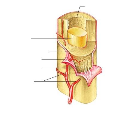
A Labelled Diagram Of Neuron with Detailed Explanations Diagram Of Neuron. A neuron is a specialized cell, primarily involved in transmitting information through electrical and chemical signals. They are found in the brain, spinal cord and the peripheral nerves. A neuron is also known as the nerve cell. The structure of a neuron varies with their shape and size and it mainly depends upon their ...
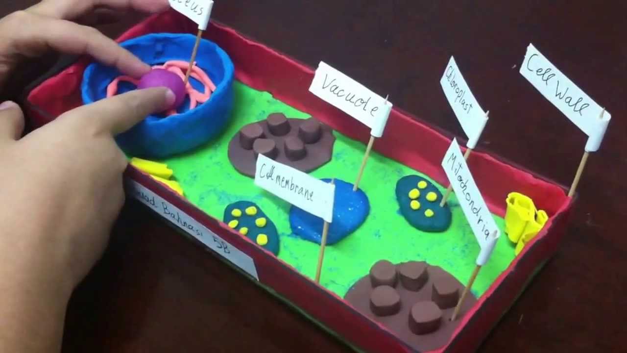
5th grade plant cell project - YouTube
Labeling a Cell Diagram | Quizlet Cell Wall This gives shape and support to the plant cell. It surrounds the cell and protects the other parts of the cell. Chloroplasts This is where the plant cell's chlorophyll is stored. This is what the plant uses to make its own food (photosynthesis). This is also what makes plant cells have a green-like color. Plant cells Are circular in shape

Labeled Volvox Diagram - Made By Creative Label
Cell: Structure and Functions (With Diagram) - Biology Discussion Eukaryotic Cells: 1. Eukaryotes are sophisticated cells with a well defined nucleus and cell organelles. 2. The cells are comparatively larger in size (10-100 μm). 3. Unicellular to multicellular in nature and evolved ~1 billion years ago. 4. The cell membrane is semipermeable and flexible. 5. These cells reproduce both asexually and sexually.

Bernadette's Life Science Blog
Cells Diagram | Science Illustration Solutions - Edrawsoft Cells Diagram Symbols Edraw software offers you lots of symbols used in cells diagram like cell structure, paramecium, squamous cell, cell division, bacteria, cell membrane, eggs, sperm, zygote, an animal cell, SARS, tobacco mosaic, adenovirus, coliphage, herpesvirus, AIDS, pollen, plant cell model, onion tissue, etc. Cells Diagram Examples
A Typical Cell, Labeled Stock Photo 112395266 : Shutterstock
Learn the parts of a cell with diagrams and cell quizzes - Kenhub For this exercise we'll start with an image of a cell diagram ready labeled. Study this and make sure that you're clear about which structure is found where. Cell diagram unlabeled It's time to label the cell yourself! As you fill in the cell structure worksheet, remember the functions of each part of the cell that you learned in the video.

HBSE I: June 2010
How to draw a nerve cell - labeled science diagrams - YouTube Download a free printable outline of this video and draw along with us: you for watching. Please su...

Hemopoiesis (formation of blood cells) - YouTube
Plant Cells: Labelled Diagram, Definitions, and Structure Plants have a rigid cell wall that surrounds the plasma membrane. The cell wall is made of cellulose and lignin, which are strong and tough compounds. Plant Cells Labelled Plastids and Chloroplasts Plants make their own food through photosynthesis. Plant cells have plastids, which animal cells don't.
Label the Cell Diagram
Human Cell Diagram, Parts, Pictures, Structure and Functions One of the few cells in the human body that lacks almost all organelles are the red blood cells. The main organelles are as follows : cell membrane endoplasmic reticulum Golgi apparatus lysosomes mitochondria nucleus perioxisomes microfilaments and microtubules Diagram of the human cell illustrating the different parts of the cell. Cell Membrane

30 Animal Cell Model Diagram And How To Understand Them - front yard landscaping ideas | Animal ...
Cell Organelles- Definition, Structure, Functions, Diagram Cell organelles are specialized entities present inside a particular type of cell that performs a specific function. There are various cell organelles, out of which, some are common in most types of cells like cell membranes, nucleus, and cytoplasm. However, some organelles are specific to one particular type of cell-like plastids and cell ...

Cell Diagram With Labels - General Wiring Diagram
Diagram of a cell membrane with labels - NIST Essential Biological FunctionsImmune response, Cell metabolism, Neurotransmission, Photosynthesis, Cell adherence, Cell growth and differentiationPotential Commercial ApplicationsDrug response monitoring, Chemical manufacturing, Biosensing, Energy conversion, Tissue engineering ... Diagram of a cell membrane with labels. Appears In. Biology in ...

Converting Diagrams
Label the cell - Teaching resources - Wordwall G5 Science. Label the Plant Cell Labelled diagram. by Koneal2. G7 Biology. Label Animal Cell Organelles Labelled diagram. by Britter. Correctly Label the Bacteria (Prokaryotic) Cell Labelled diagram. by Bronwyn12. Label Plant and Animal Cell Labelled diagram.

Animal Cell Royalty Free Stock Photo - Image: 29859935
Anatomy – Page 58 – Graph Diagram
Wiring Diagram Database: Human Cell Diagram To Label

34 Human Cell Diagram To Label - Labels For Your Ideas
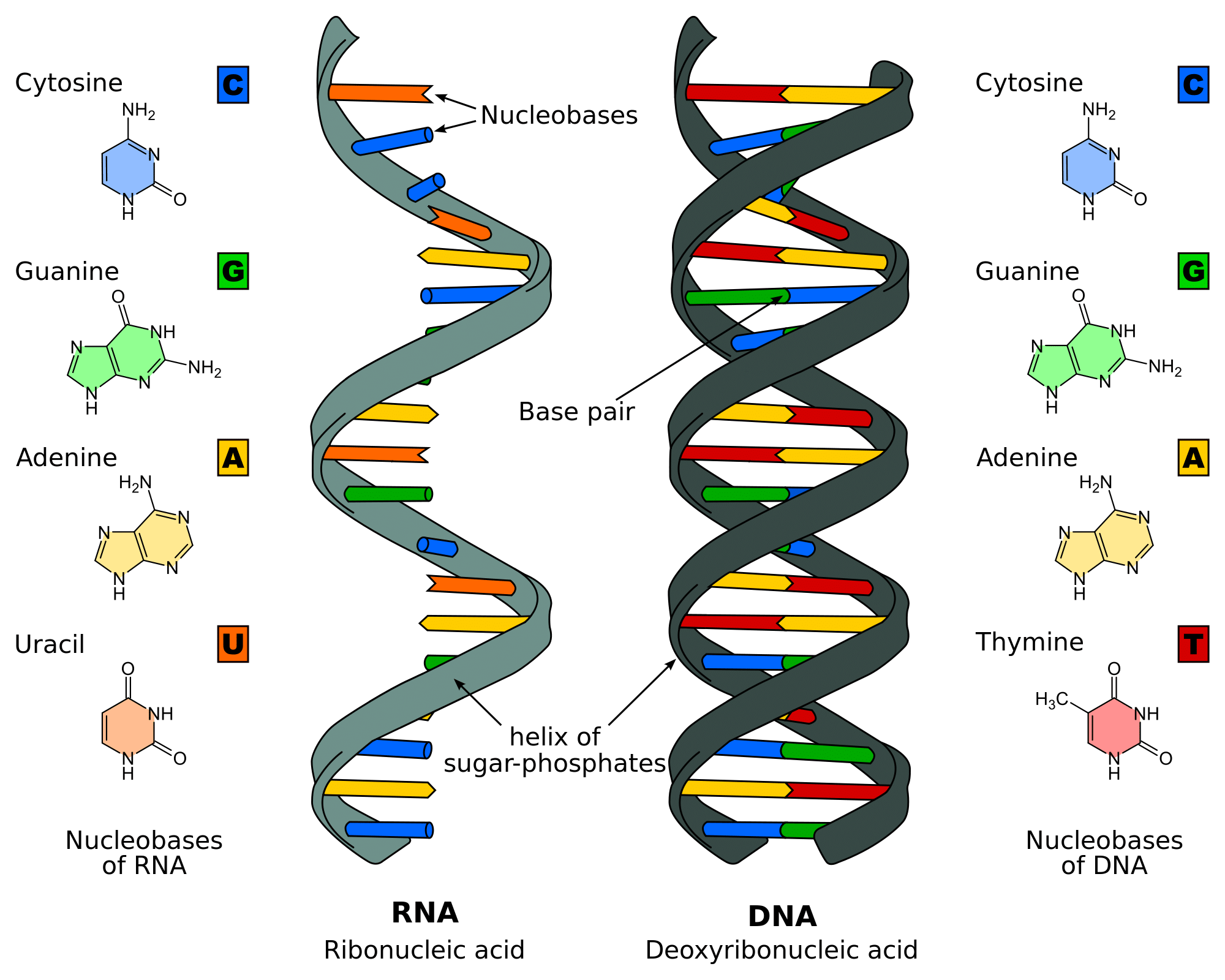
DNA Replication - Structure - Stages of Replication - TeachMePhyiology
Epithelia: The Histology Guide
Post a Comment for "43 diagram of a cell with labels"