40 human skull diagram with labels
jose-santos.com › rjmegyj › cadaver-anatomy-picturescadaver anatomy pictures Nerve ( 12 cards ) 2022-05-11 47, TeachMeAnatomy is a comprehensive anatomy encyclopaedia presented a!, Ricki Lewis head with skull in ghost effect, side view is an old science, having its in! Them for you stock pictures, cadaver anatomy pictures photos & amp ; images structures—muscles, tendons, and human... Free Printable Label A Skeleton Worksheet For Kids This label a skeleton worksheet for kids is perfect for children learning about the human body or for an educational Halloween activity!. Over the past few days, we've been teaching all about the human body. We've used these human body worksheets, did this easy skeleton handprint craft, made this cool germ blow painting art, and told stories with these 5 senses puppets.
Free Skeletal System Worksheets and Printables We have created the Human Body Systems Labeling and Diagramming Worksheet as an instant download. This includes fill-in-the-blanks for your student to label the main bones on the body. Label the Skeleton Activity - This worksheet walks your children through labeling a skeleton. You'll label the main bones of the body.

Human skull diagram with labels
Printable Unlabeled Blank Skeleton Diagram - Muro Fornew Printable human skeleton diagram labeled unlabeled and . The parts of the skeleton have been labeled. this flexible inner framework supports all other parts and tissues, which would collapse without skeletal reinforcement. In this worksheet, we are going to review some of the major bones within the body. Types of Bones in the Human Body: Skeletal System Labeled Diagram and ... Skeletal System Types of Bones: Labeled diagram of flat bones which include the bones of the rib cage and parts of the skull. F = Flat = Fort (Rib Cage & Skull) Example Flat Bones The average adult has 36 flat bones. Which bones are considered flat bones? Frontal Bone - Location, Functions, Anatomy, & Diagram Plays a major role in a person's appearance. Frontal Bone Anatomy It is a bowl-shaped bone, comprising three parts: the squamous part, the orbital part, and the nasal part. Frontal Bone Anatomy Labeled 1. Squamous Part It is the largest area of the bone, encircling the forehead. Its external side is flat, but the internal side is concave.
Human skull diagram with labels. Sternum - Anatomy, Parts, Location, Functions, & Diagram Sternum, commonly called breastbone, is a long, flat bone located in the midline of the chest. The word 'sternum' has been derived from the ancient Greek word ' sternon ', meaning 'chest'. The bone covers and protects the thoracic organs, such as heart, and lungs from any external shock. Human Skull Anatomy, Bones & Functions - Study.com Skull Diagram Here is a skull diagram that shows the skull labeled from the side view as well as from the front view. Front view of human skull labeled Side view of human skull labeled The Skull's... › stock-photo › female-anatomy-diagramFemale Anatomy Diagram Stock Photos and Images - Alamy Find the perfect female anatomy diagram stock photo. Huge collection, amazing choice, 100+ million high quality, affordable RF and RM images. No need to register, buy now! Skull: Anatomy, structure, bones, quizzes | Kenhub The human skull consists of 22 bones (or 29, including the inner ear bones and hyoid bone) which are mostly connected together by ossified joints, so called sutures. The skull is divided into the braincase ( neurocr anium) and the facial skeleton ( viscerocranium ).
› en › e-AnatomyAnatomical diagrams of the brain - e-Anatomy - IMAIOS Sep 13, 2021 · The study of the arterial supply of blood to the brain is facilitated by a diagram showing the cerebral arterial vascular areas in lateral and medial views and axial and coronal section and by diagrams of arteries forming the Willis' circle (internal and vertebral carotid arteries, basilar artery, anterior and posterior communicating arteries ... Chart of Major Muscles on the Front of the Body with Labels The coracobrachialis is the smallest of the three muscles that attach to the coracoid process of the scapula. (The other two muscles that attach here are the pectoralis minor and the short head of the biceps brachii.) It is situated at the upper and medial part of the arm. It is supplied by the musculocutaneous nerve. en.wikipedia.org › wiki › File:Human_skeleton_frontFile:Human skeleton front en.svg - Wikipedia This image was previously a featured picture, but community consensus determined that it no longer meets our featured-picture criteria.If you have a high-quality image that you believe meets the criteria, be sure to upload it, using the proper free-license tag, then add it to a relevant article and nominate it. Anatomy of the spinal cord - e-Anatomy - IMAIOS This atlas of human anatomy describes the spinal cord through 18 anatomical diagrams with 270 anatomical structures labeled. It was designed particularly for physiotherapists, osteopaths, rheumatologists, neurosurgeons, orthopedic surgeons and general practitioners, especially for the study and understanding of medullary diseases.
A Student's Guide to Learning the Human Bones A Student's Guide to Learning the Human Bones. Shannan Muskopf October 30, 2021. Most anatomy classes include a unit on the human skeleton. I have several disarticulated human skeletons and quite a few plastic models. Students use them to identify bones and structures on the bones. The activity as a way for students to examine diagrams on ... Anatomy of the spine and back - e-Anatomy - IMAIOS Anatomical diagrams of the spine and back. This human anatomy module is composed of diagrams, illustrations and 3D views of the back, cervical, thoracic and lumbar spinal areas as well as the various vertebrae. It contains the osteology, arthrology and myology of the spine and back. It is particularly interesting for physiotherapists ... Leg Bones Anatomy, Names & Diagram - Study.com The major leg bones in the body are: Femur: the upper leg in both legs. Patella: the kneecap in both legs. Tibia: the larger of the two bones in the lower leg, which supports the body. Fibula: the ... Anatomy Of Human Body Parts Diagram - 9 images - Genentech [Anatomy Of Human Body Parts Diagram] - 9 images - phenomia symptoms hrf, brain anatomy internal structures medical art library, ... Basic Human Skeleton Diagram. Gallery of Anatomy Of Human Body Parts Diagram. Fahrer Jobs Kempten Dover De It Jobs Steve Jobs Krankheit Pwc Hr 8 Stunden Job Berlin Jobs Hannover Französisch Bundeswehr Zivil Job ...

Unlabelled Respiratory System Clip Art at Clker.com - vector clip art online, royalty free ...
Human Spine Diagram Labeled - vertebral column labeling purposegames ... Human Spine Diagram Labeled - 9 images - labeled picture of the nervous system nervous system, printable skeleton diagram print to label skeletal, ... We take on this nice of Human Spine Diagram Labeled graphic could possibly be the most trending topic in imitation of we share it in google plus or facebook.
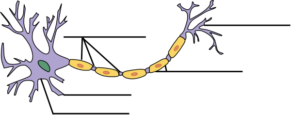
Label The Neuron Clip Art at Clker.com - vector clip art online, royalty free & public domain
Human Muscles Diagram - Ochinang72998 This diagram depicts human muscle system diagram with parts and labels. Today s blog is all about muscle everybody has loads of muscles in. Posted in anatomy, bones, muscles | tagged body skeleton, human muscle diagram, human muscles, human. There are around 650 skeletal muscles within the typical human body.
Post a Comment for "40 human skull diagram with labels"