39 onion cells under microscope with labels
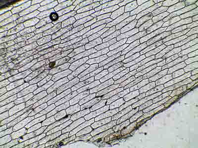
Microscope Onion Cell Diagram - Wiring Schematic Online Sketch the onion peel cell as seen under the microscope label the. The cheek epithelium cell is the only one that has centrioles the barrel shaped organelle that is responsible for helping organize chromosomes during cell division. Preparation and viewing of onion cells expected time for completion. Observe an onion cell under the microscope. Epidermal onion cells under a microscope. Plant cells appear polygonal ... Observing onion cells under the microscope. For this microscope experiment, the thin membrane will be used to observe the cells. An easy beginner experiment. Jessica Williams. Ideas for Work. Similar ideas popular now. Applied Science. Subjects. Physical Science. Technology.
Plant Cell Under Microscope 40X Labeled : 1 - Chloroplast and cell wall ... The different images below were taken with two different types of microscopes. 1.can only turn fine adjustment 2.draw one row of cells across the middle 3.label the chloroplasts and cell wall. When using the microscope always start by focusing under low power and working your way up to high power.

Onion cells under microscope with labels
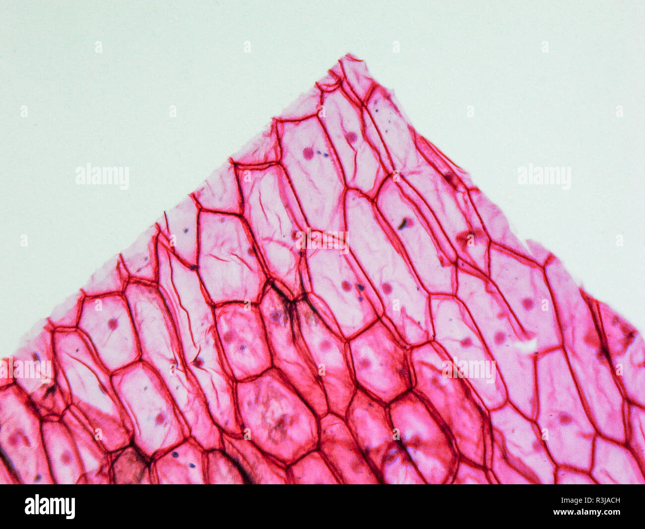
How to observe onion cells under a microscope? - JacAnswers How to observe onion cells under a microscope? Gently lay a microscopic cover slip on the membrane and press it down gently using a needle to remove air bubbles. Touch a blotting paper on one side of the slide to drain excess iodine/water solution, Place the slide on the microscope stage under low power to observe. Onion Epidermal Cell Labeled Diagram - schematron.org Nov 15, 2018 · Draw a labelled diagram of an onion epidermal cell seen under the microscope. ( 4 marks) e The onion epidermal cells are not green in colour because they lack. The epidermal cells of onions provide a protective layer against viruses and fungi that may harm the sensitive tissues. Onion Cells Microscope Stock Photos and Images - Alamy Onion cells under the microscope. Garden onion, Bulb Onion, Common Onion (Allium cepa), cell tissue of a garden onion with dyed chromosomes, light microscopy, x 120, Germany. Onion Cells under the Microscope. Onion skin cells under the microscope, horizontal field of view is about 0.61 mm. Detailed view of the cells of a red onion as seen ...
Onion cells under microscope with labels. Observing Onion Cells Under The Microscope Afterwards, carefully mount the prepared and stained onion cell slide onto the microscope stage. Make sure that the cover slip is perfectly aligned with the microscope slide, and that any excess stain has been wiped off. Secure the slide on the stage using the stage clips. PDF Onion Cell Lab - somewaresinmaine.com Research Biology Onion Cell Lab page 1 of 3 Onion Cell Lab After you have completed the rest of this lab come back to this cover page DRAW & LABEL AN ONION CELL WITH ALL THE PARTS / ORGANELLES YOU OBSERVE UNDER 40X. Purpose: To observe and identify major plant cell structures and to relate the structure of the cell to its function. Materials: 1 ... Cell Onion Under Labeled Microscope 4250 microscope EUR-BB-4250 1 2 Microscopic slides, 50 pcs 64691-00 1 3 Cover glasses 18x18 mm, 50 pcs YOU WILL NEED: An onion, a slide and cover slip, a cotton bud, some food colouring, a plate to Label the cell wall, cell membrane, and cytoplasm Label the diagrams and fill in the chart on your post-lab question sheet This work is licensed under a Creative Commons Attribution-NonCommercial ... Onion Epidermis - kuensting.org Onion epidermal cells, iodine stain, 400X. The nucleus of an onion epidermal cell, 1000X magnification. ...
Onion Cell Diagram Labeled Pdf Copy - thesource2.metro Just invest tiny become old to gain access to this on-line broadcast onion cell diagram labeled pdf as skillfully as review them wherever you are now. Grade 8 Intermediate-level Science Test June 2021 (v202) … diagram below and on your knowledge of science. The diagram represents a potato plant. Several plant structures are labeled. Looking at the Structure of Cells in the Microscope Both types of light microscopy are widely used to visualize living cells. Figure 9-7 Two ways to obtain contrast in light microscopy. (A) The stained portions of the cell reduce the amplitude of light waves of particular wavelengths passing through them. A colored image of the cell is thereby obtained that is visible in the ordinary way. (more...) Microscopy, size and magnification - Microscopy, size and ... - BBC Place cells on a microscope slide. Add a drop of water or iodine (a chemical stain). Lower a coverslip onto the onion cells using forceps or a mounted needle. This needs to be done gently to... Blog, She Wrote - Embracing the Independent & Authentic Nature of ... Blog, She Wrote - Embracing the Independent & Authentic Nature of ...
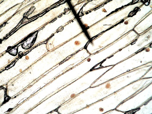
Onion Root Tip Mitosis - Stages, Experiment and Results · Cover the sample (root tip) with a coverslip and gently press the coverslip down, then examine the slide under the microscope starting with low magnification * For this experiment, a properly prepared slide should appear light pink due to the stain to almost colorless. * Unused roots can be stored in 70 percent alcohol. Results Onion Cells Under a Microscope - Requirements/Preparation/Observation Add a drop of iodine solution on the onion membrane (or methylene blue) Gently lay a microscopic cover slip on the membrane and press it down gently using a needle to remove air bubbles. Touch a blotting paper on one side of the slide to drain excess iodine/water solution, Place the slide on the microscope stage under low power to observe. Animal Cell Diagram Under Microscope Labeled - ACTUINDE Animal Cell Diagram Under Microscope Labeled Sunday, April 18th 2021. | Diagram Animal Cell Diagram Under Microscope. Function cell does in the body dictate the change and adaptation done by cell. When observing onion cells, there is the Cell Surface Membrane which is present in all living cells. DOC Plant and Animal Cells Microscope Lab - Hillsboro City Schools Make a drawing of one onion cell, labeling all of its parts as you observe them. (At minimum you should observe the nucleus, cell wall, and cytoplasm.) Cheek cells 1. To view cheek cells, gently scrape the inside lining of your cheek with a toothpick. DO NOT GOUGE THE INSIDE OF YOUR CHEEK! (We will observe blood cells in a future lab!!) 2.
Cryogenic scanning electron micrograph of the interface | Open-i
The following diagram shows cells of onion peel label class ... - Vedantu 115.2k + views. Hint: The diagrams mentioned above are the internal structure of an onion peel and human cheek cells. In order to label them, we need to understand its anatomy and know about various structures present in it. Onion peel is an example of a plant cell whereas a human cheek cell is an example of an animal cell. Complete answer:

Cell Biology Project - Parts of a cell - Quatr.us Study Guides
Plant Cell Under Microscope Labeled 40X : Young Root 2 Of Broad Bean ... Cells and viewing them under the microscope. A small square of a red onion skin (membrane) was observed under a microscope at high power (x40) magnification. (iv) describe how you applied the stain. They must draw and label the nucleus, cell membrane set up your microscope, place the onion root slide on the stage and focus on low (40x) power.

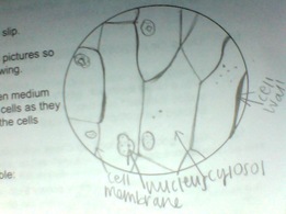
Sketch the onion peel cell as seen under the microscope. Label the parts such as the cell wall ...
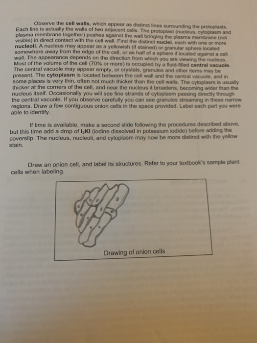
PDF Onion Cells - Investigation - Exploring Nature 5. Observe the onion tissue under the microscope at 4x, 10x and 40x with lots of light (open diaphragm). Then slowly close the diaphragm while observing the image to find the best light for seeing cellular details. 6. Draw a section of onion skin cells at 10x magnification. Then switch to 40x and draw one cell and label it. Questions: 1.
Onion Cells Microscope High Resolution Stock Photography and Images - Alamy
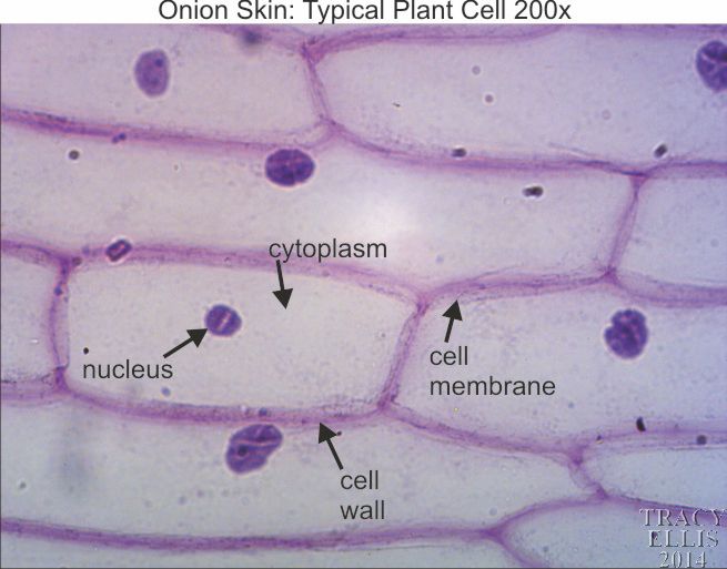
DOC The Onion Cell Lab - chsd.us Onion tissue provides excellent cells to study under the microscope. The main cell structures are easy to see when viewed with the microscope at medium power. For example, you will observe a large circular . nucleus. in each cell, which contains the genetic material for the cell. In each nucleus, are round bodies called . nucleoli

Root Tip Of Onion And Mitosis Cell In The Root Tip Of Onion Under A Microscope Stock Photo ...
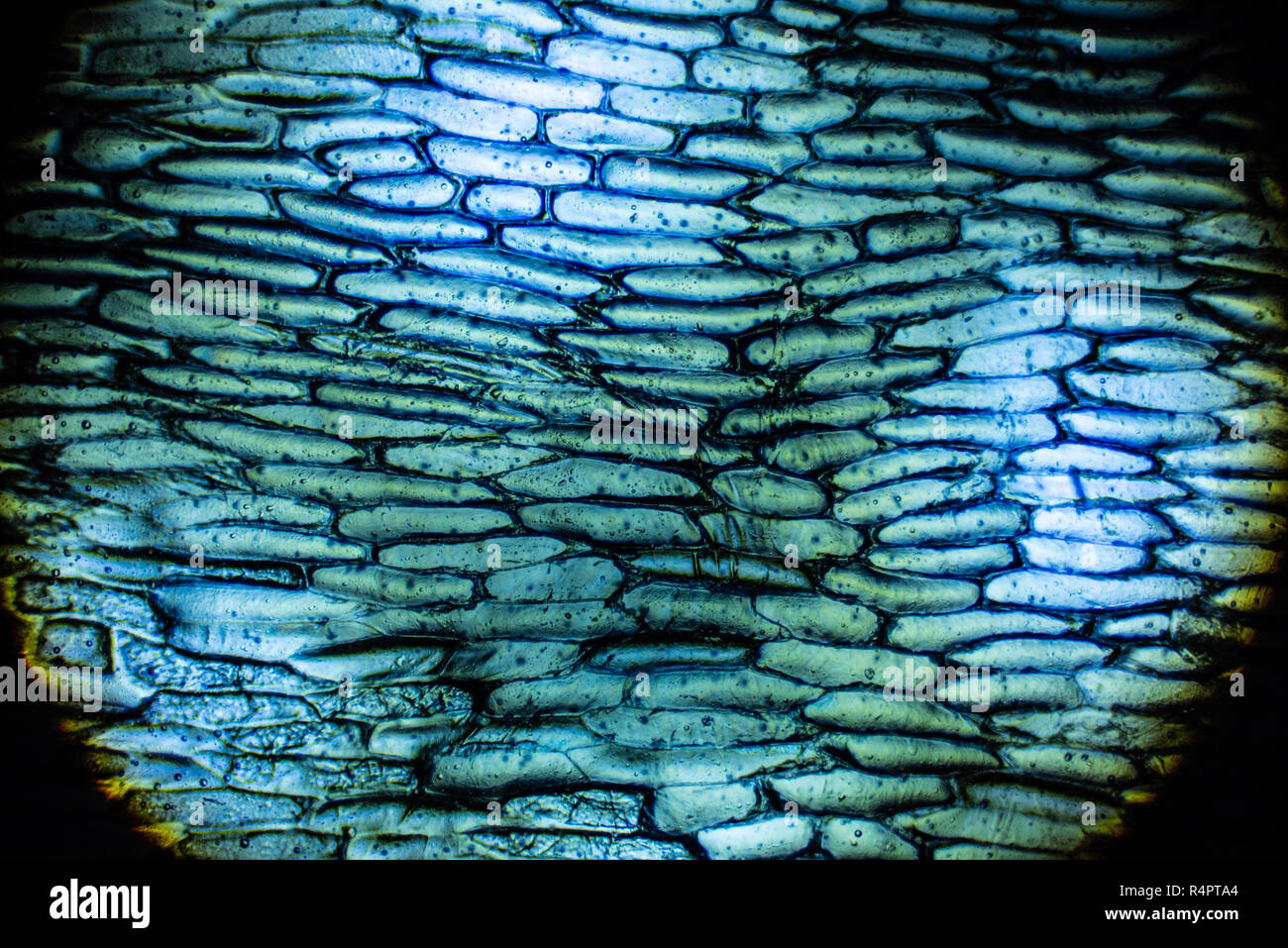
Onion Cells Microscope Stock Photos and Images - Alamy Onion cells under the microscope. Garden onion, Bulb Onion, Common Onion (Allium cepa), cell tissue of a garden onion with dyed chromosomes, light microscopy, x 120, Germany. Onion Cells under the Microscope. Onion skin cells under the microscope, horizontal field of view is about 0.61 mm. Detailed view of the cells of a red onion as seen ...

Biology Pictures: Onion Cells under Microscope | Biology, Cell biology, Picture
Onion Epidermal Cell Labeled Diagram - schematron.org Nov 15, 2018 · Draw a labelled diagram of an onion epidermal cell seen under the microscope. ( 4 marks) e The onion epidermal cells are not green in colour because they lack. The epidermal cells of onions provide a protective layer against viruses and fungi that may harm the sensitive tissues.
Onion Cells High Resolution Stock Photography and Images - Alamy
How to observe onion cells under a microscope? - JacAnswers How to observe onion cells under a microscope? Gently lay a microscopic cover slip on the membrane and press it down gently using a needle to remove air bubbles. Touch a blotting paper on one side of the slide to drain excess iodine/water solution, Place the slide on the microscope stage under low power to observe.
My Personal Experience - SusannaLaRochellemicroscopy
Cyanamide mode of action during inhibition of onion ( Allium cepa L.) root growth involves ...
Microscope Onion Cell Labeled - Micropedia
General Biology Microscopic Specimen Images & Photographs

Fanos' MCB Blog
Microscope Onion Cell Labeled - Micropedia
Biology Pictures: Onion Cells under Microscope

The Microscope

Onion Cell Under Microscope 40x Labeled - Micropedia

Life Science Unit - WELCOME TO MR.FLEMING SCIENCE
Post a Comment for "39 onion cells under microscope with labels"